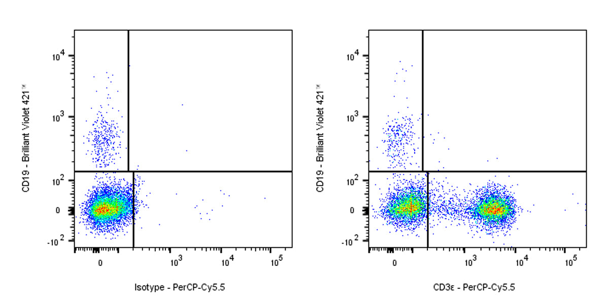
Flow cytometric analysis of Human CD3ε expression on human peripheral blood leukocytes. Human peripheral blood leukocytes were stained with Brilliant Violet 421™ Mouse Anti-Human CD19 antibody and either PerCP-Cy5.5 Mouse IgG2a Isotype Control (Left panel) or SDT PerCP-Cy5.5 Mouse Anti-Human CD3ε Antibody (Right panel) at 5 μl/test treated with True-Stain Monocyte Blocker™. Total viable cells, as determined by Fixable Viability Dye 515 (S0D0013), were used for analysis. Flow cytometry and data analysis were performed using BD FACSymphony™ A1 and FlowJo™ software.
PerCP-Cy5.5 Mouse Anti-Human CD3ε Antibody (HIT3a)
PerCP-Cy5.5 Mouse Anti-Human CD3ε Antibody (HIT3a)
Price:
Regular price
$146 USD
Regular price
Sale price
$146 USD
Unit price
per
For shipping services or bulk orders, you may request a quotation.
Secure checkout with

View full details
Product Details
Product Details
Product Specification
| Synonyms | T-cell surface glycoprotein CD3 epsilon chain; T-cell surface antigen T3/Leu-4 epsilon chain; T3E; CD3E |
| Location | Cell membrane |
| Accession | P07766 |
| Clone Number | HIT3a |
| Antibody Type | Mouse mAb |
| Isotype | IgG2a |
| Application | FCM |
| Reactivity | Hu |
| Purification | Protein A |
| Concentration | 0.2 mg/ml |
| Conjugation | PerCP-Cy5.5 |
| Physical Appearance | Liquid |
| Storage Buffer | PBS, 1% BSA, 0.3% Proclin 300 |
| Stability & Storage | 12 months from date of receipt / reconstitution, 2 to 8 °C as supplied |
Dilution
| application | dilution | species |
| FCM | 5μl per million cells in 100μl volume | Hu |
Background
CD3ε is a subunit of the CD3 complex, which is a critical component of the T-cell receptor (TCR) complex on T cells. Together with CD3γ, CD3δ, and CD3ζ, it forms the TCR-CD3 complex that plays a vital role in coupling antigen recognition to intracellular signaling pathways. This complex is essential for T cell activation, proliferation, and differentiation. CD3ε contains an extracellular immunoglobulin domain, a transmembrane region, and a cytoplasmic tail with an immunoreceptor tyrosine-based activation motif (ITAM). Upon antigen recognition, the ITAMs in CD3ε are phosphorylated, initiating downstream signaling cascades that lead to T cell activation. Additionally, CD3ε interacts with other signaling molecules like Lck and Nck through its unique motifs, further modulating T cell responses. Its importance in T cell function makes CD3ε a target for immunotherapies, including cancer treatments and autoimmune disease management.
Picture
Picture
FC