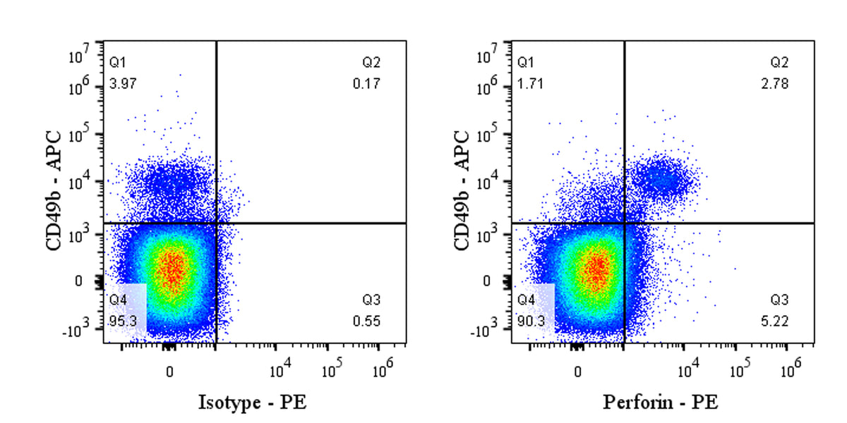
Flow cytometric analysis of Mouse Perforin expression in C57BL/6 mouse splenocytes. C57BL/6 mouse splenocytes were fixed and permeabilized with Foxp3 / Transcription Factor Staining Buffer Set. The cells were then washed and stained in Perm/Wash™ Buffer with APC Rat Anti-Mouse CD49b Antibody and with either PE Rat IgG2a, κ Isotype Control or SDT PE Rat Anti-Mouse Perforin Antibody at 5 μl/test. Total viable cells, as determined by Fixable Viability Dye 452 (S0B88801), were used for analysis. Flow cytometry and data analysis were performed using BD FACSymphony™ A1 and FlowJo™ software.
PE Rat Anti-Mouse Perforin Antibody (S-R644)
PE Rat Anti-Mouse Perforin Antibody (S-R644)
Price:
Regular price
$85 USD
Regular price
Sale price
$85 USD
Unit price
per
For shipping services or bulk orders, you may request a quotation.
Secure checkout with

View full details
Product Details
Product Details
Product Specification
| Host | Rat |
| Antigen | Perforin |
| Synonyms | Perforin-1; P1; Cytolysin; Lymphocyte pore-forming protein; Pfp; Prf1 |
| Location | Secreted, Cell membrane |
| Accession | P10820 |
| Clone Number | S-R644 |
| Antibody Type | Rat mAb |
| Isotype | IgG2a,k |
| Application | ICFCM |
| Reactivity | Ms |
| Positive Sample | C57BL/6 mouse splenocytes |
| Purification | Protein G |
| Concentration | 0.2 mg/ml |
| Conjugation | PE |
| Physical Appearance | Liquid |
| Storage Buffer | PBS, 1% BSA, 0.09% sodium azide |
| Stability & Storage | 12 months from date of receipt / reconstitution, 2 to 8 °C as supplied |
Dilution
| application | dilution | species |
| ICFCM | 1μg per million cells in 100μl volume | Ms |
Background
Perforin is a crucial protein involved in the immune response, primarily produced by cytotoxic T lymphocytes and natural killer cells. It functions by forming pores in the membranes of target cells, such as virus-infected or cancerous cells. Once released from immune cells, perforin molecules aggregate and insert into the target cell membrane, creating channels that disrupt the cell's osmotic balance, leading to cell lysis and death. This mechanism is essential for eliminating harmful cells and maintaining the body's overall health.
Picture
Picture
FC