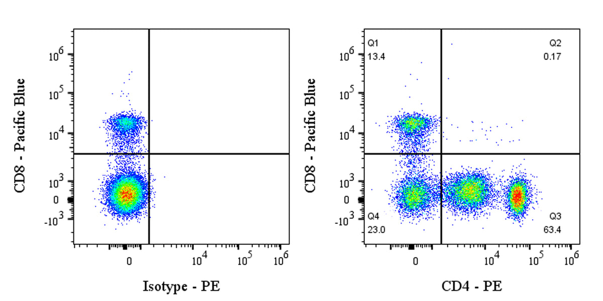
Flow cytometric analysis of Human CD4 expression on human PBMCs (human peripheral blood mononuclear cells). Human PBMCs were stained with Pacific Blue Mouse Anti-Human CD8 Antibody (S0B8156) and either PE Rabbit IgG Isotype Control (left panel) or SDT PE Rabbit Anti-Human CD4 Antibody (right panel) at 5 μl/test. Flow cytometry and data analysis were performed using Agilent NovoCyte Quanteon and FlowJo™ software.
PE Rabbit Anti-Human CD4 Antibody (13B8.2)
PE Rabbit Anti-Human CD4 Antibody (13B8.2)
Price:
Regular price
$95 USD
Regular price
Sale price
$95 USD
Unit price
per
For shipping services or bulk orders, you may request a quotation.
Secure checkout with

View full details
Product Details
Product Details
Product Specification
| Host | Rabbit |
| Antigen | CD4 |
| Location | Cell membrane, Endoplasmic reticulum |
| Accession | P01730 |
| Clone Number | 13B8.2 |
| Antibody Type | Recombinant mAb |
| Isotype | IgG |
| Application | FCM |
| Reactivity | Hu |
| Positive Sample | Human PBMC |
| Purification | Protein A |
| Concentration | 0.2 mg/ml |
| Conjugation | PE |
| Physical Appearance | Liquid |
| Storage Buffer | PBS, 1% BSA, 0.09% sodium azide |
| Stability & Storage | 12 months from date of receipt / reconstitution, 2 to 8 °C as supplied |
Dilution
| application | dilution | species |
| FCM | 5μl per million cells in 100μl volume | Hu |
Background
CD4 is a glycoprotein found on the surface of certain immune cells, such as helper T cells, monocytes, macrophages, and dendritic cells. It is a monomeric type I transmembrane molecule composed of four immunoglobulin-like domains (D1 to D4) and encoded by a single gene. As a co-receptor for the T-cell receptor (TCR), CD4 plays a crucial role in the immune response by binding to the MHC class II molecules on antigen-presenting cells, which helps T cells recognize and respond to foreign antigens. The interaction between CD4 and MHC class II molecules also activates downstream signaling pathways, leading to the activation of transcription factors and promoting T cell activation. Additionally, CD4 is significant in HIV infection, as the virus binds to CD4 to enter host cells.
Picture
Picture
FC