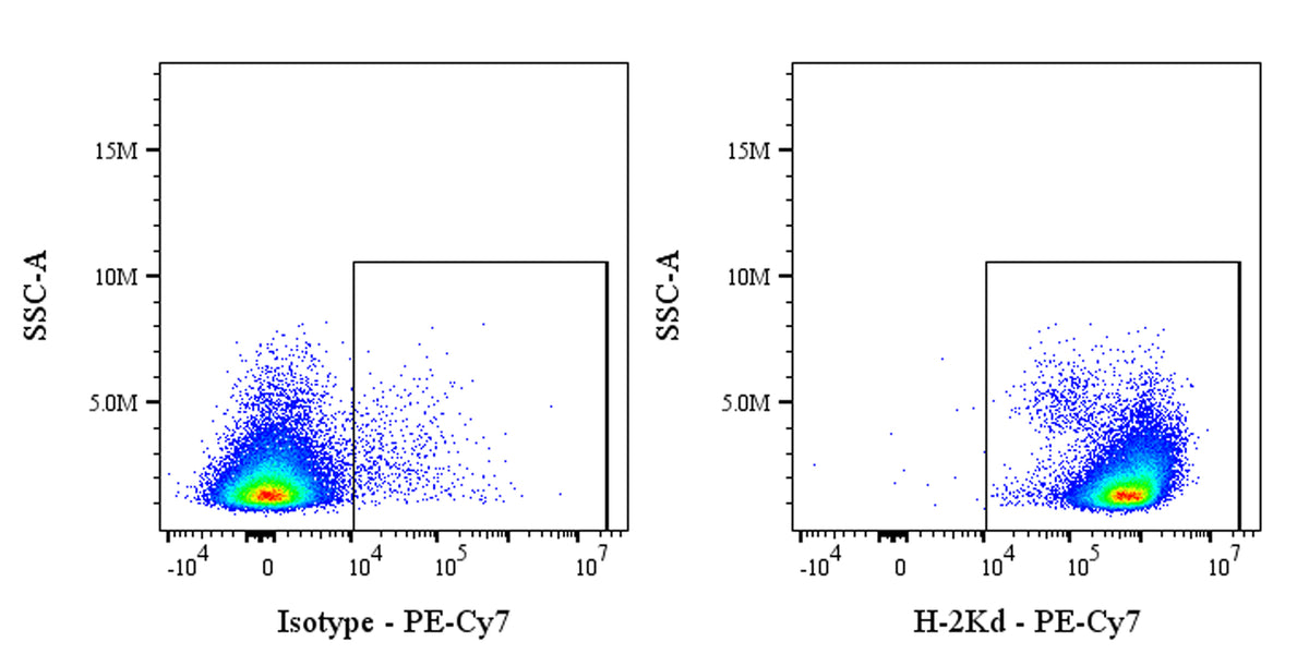
Flow cytometric analysis of Mouse H-2Kd expression on BALB/c mouse splenocytes. BALB/c mouse splenocytes were stained with PE-Cy7 Mouse IgG2a, κ Isotype Control (left panel) or SDT PE-Cy7 Mouse Anti-Mouse H-2Kd Antibody (right panel) at 1.25 μl/test treated with True-Stain Monocyte Blocker™. Flow cytometry and data analysis were performed using Agilent NovoCyte Quanteon and FlowJo™ software.
PE-Cy7 Mouse Anti-Mouse H-2Kd Antibody (SF1-1.1)
PE-Cy7 Mouse Anti-Mouse H-2Kd Antibody (SF1-1.1)
Price:
Regular price
$160 USD
Regular price
Sale price
$160 USD
Unit price
per
For shipping services or bulk orders, you may request a quotation.
Secure checkout with

View full details
Product Details
Product Details
Product Specification
| Host | Mouse |
| Antigen | H-2Kd |
| Synonyms | H-2 class I histocompatibility antigen, K-D alpha chain; H-2K(D); H2-K; H2-K1 |
| Location | Membrane |
| Accession | P01902 |
| Clone Number | SF1-1.1 |
| Antibody Type | Mouse mAb |
| Isotype | IgG2a,k |
| Application | FCM |
| Reactivity | Ms |
| Positive Sample | BALB/c mouse splenocytes |
| Purification | Protein A |
| Concentration | 0.2 mg/ml |
| Conjugation | PE-Cy7 |
| Physical Appearance | Liquid |
| Storage Buffer | PBS, 1% BSA, 0.09% sodium azide |
| Stability & Storage | 12 months from date of receipt / reconstitution, 2 to 8 °C as supplied |
Dilution
| application | dilution | species |
| FCM | 0.25μg per million cells in 100μl volume | Ms |
Background
H-2Kd is a murine MHC class I molecule expressed by BALB/c mice that presents endogenous 9-mer peptides—anchored by Tyr at position 2 and a hydrophobic residue at position 9—to CD8⁺ cytotoxic T cells, thereby initiating adaptive immune responses; its 2.6-Å crystal structure reveals a peptide-binding groove with five deep pockets (A–F) that impose an allele-specific motif, and it pairs non-covalently with β2-microglobulin for stability.
Picture
Picture
FC