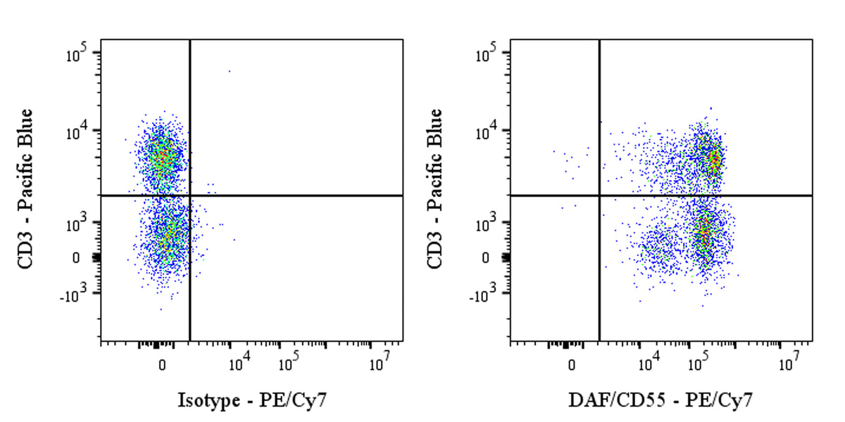
Flow cytometric analysis of human CD55 expression on human peripheral blood cells. Human peripheral blood cells were stained with Pacific Blue Mouse Anti-Human CD3 Antibody (S0B5475) and either PE-Cy7 Mouse IgG1, k Isotype Control (left panel) or PE-Cy7 Mouse Anti-Human CD55 Antibody (right panel) at 2 μl/test treated with True-Stain Monocyte Blocker™. Total viable cells, as determined by Fixable Viability Dye 515 (S0B88804), were used for analysis. Flow cytometry and data analysis were performed using Agilent NovoCyte Quanteon and FlowJo™ software.
PE-Cy7 Mouse Anti-Human DAF/CD55 Antibody (S-628-48)
PE-Cy7 Mouse Anti-Human DAF/CD55 Antibody (S-628-48)
Price:
Regular price
$205 USD
Regular price
Sale price
$205 USD
Unit price
per
For shipping services or bulk orders, you may request a quotation.
Secure checkout with

View full details
Product Details
Product Details
Product Specification
| Host | Mouse |
| Antigen | DAF/CD55 |
| Synonyms | Complement decay-accelerating factor; CR |
| Immunogen | Recombinant Protein |
| Location | Secreted, Cell membrane |
| Accession | P08174 |
| Clone Number | S-628-48 |
| Antibody Type | Mouse mAb |
| Isotype | IgG1,k |
| Isotype Control | S0B80046 |
| Application | FCM |
| Reactivity | Hu |
| Positive Sample | Human peripheral blood cells |
| Purification | Protein G |
| Concentration | 0.1 mg/ml |
| Conjugation | PE-Cy7 |
| Physical Appearance | Liquid |
| Storage Buffer | PBS, 1% BSA, 0.09% sodium azide |
| Stability & Storage | 12 months from date of receipt / reconstitution, 2 to 8 °C as supplied |
Dilution
| application | dilution | species |
| FCM | 2μl per million cells in 100μl volume | Hu |
Background
Decay-accelerating factor (DAF, also known as CD55) is a complement regulatory protein that plays a crucial role in protecting cells from complement-mediated attack. It is a membrane lipid microdomain-associated, GPI-anchored protein that accelerates the decay of C3 and C5 convertases, which are essential for the downstream formation of the membrane attack complex (MAC) in the complement cascade. This action helps prevent excessive complement activation and subsequent cell damage. CD55 is involved in various physiological and pathological processes, including immune regulation, cancer progression, and tissue protection. In cancer, CD55 can promote malignant transformation, cell survival, angiogenesis, and inhibit apoptosis through intracellular signaling pathways such as JNK, JAK/STAT, MAPK/NF-κB, and LCK. Additionally, it is enriched in the cancer stem cell niche and can be induced by chemotherapeutics and hypoxic environments.
Picture
Picture
FC