WB result of PDHA1 Recombinant Rabbit mAb
Primary antibody: PDHA1 Recombinant Rabbit mAb at 1/1000 dilution
Lane 1: HeLa whole cell lysate 25 μg
Lane 2: 293T whole cell lysate 25 μg
Lane 3: Hep G2 whole cell lysate 25 μg
Lane 4: MOLT4 whole cell lysate 25 μg
Lane 5: A-431 whole cell lysate 25 μg
Secondary antibody: Goat Anti-Rabbit IgG (H+L), HRP conjugated at 1/10000 dilution
Predicted MW: 43 kDa
Observed MW: 40 kDa
PDHA1 Recombinant Rabbit mAb
PDHA1 Recombinant Rabbit mAb
Price:
Regular price
$100 USD
Regular price
Sale price
$100 USD
Unit price
per
For shipping services or bulk orders, you may request a quotation.
Secure checkout with

View full details
Product Details
Product Details
Product Specification
| Host | Rabbit |
| Antigen | PDHA1 |
| Location | Mitochondrion |
| Accession | P08559 |
| Antibody Type | Recombinant mAb |
| Isotype | IgG |
| Application | WB, ICC |
| Reactivity | Hu, Ms, Rt |
| Positive Sample | HeLa, 293T, HepG2, MOLT4, A-431, NIH/3T3, Neuro-2a, mouse brain, mouse liver, PC-12, rat liver, rat brain |
| Purification | Protein A |
| Concentration | 0.4 mg/ml |
| Conjugation | Unconjugated |
| Physical Appearance | Liquid |
| Storage Buffer | PBS, 40% Glycerol, 0.05% BSA, 0.02% sodium azide |
| Stability & Storage | 12 months from date of receipt / reconstitution, -20 °C as supplied |
Dilution
| application | dilution | species |
| WB | 1:1000 | Hu, Ms, Rt |
| ICC | 1:200 | Hu |
Background
PDHA1, or pyruvate dehydrogenase E1 subunit alpha 1, is the catalytic alpha subunit of the mitochondrial pyruvate dehydrogenase (PDH) complex. It forms a heterotetrameric E1 enzyme together with the PDHB subunit and contains the active site that converts pyruvate into acetyl-CoA and CO₂, linking glycolysis to the TCA cycle. PDHA1 is a nuclear-encoded mitochondrial protein of about 43 kDa and is widely expressed in most tissues. Its activity is tightly regulated by reversible phosphorylation: PDH kinases phosphorylate specific serine residues to inhibit it, while PDH phosphatases dephosphorylate it to restore activity. PDHA1 dysfunction or mutation is associated with pyruvate dehydrogenase E1-alpha deficiency and X-linked Leigh syndrome, and it is also studied in cancer metabolism, aging, and other metabolic disorders.
Picture
Picture
Western Blot
WB result of PDHA1 Recombinant Rabbit mAb
Primary antibody: PDHA1 Recombinant Rabbit mAb at 1/1000 dilution
Lane 1: NIH/3T3 whole cell lysate 25 μg
Lane 2: Neuro-2a whole cell lysate 25 μg
Lane 3: mouse brain lysate 25 μg
Lane 4: mouse liver lysate 25 μg
Secondary antibody: Goat Anti-Rabbit IgG (H+L), HRP conjugated at 1/10000 dilution
Predicted MW: 43 kDa
Observed MW: 40 kDa
WB result of PDHA1 Recombinant Rabbit mAb
Primary antibody: PDHA1 Recombinant Rabbit mAb at 1/1000 dilution
Lane 1: PC-12 whole cell lysate 25 μg
Lane 2: rat brain lysate 25 μg
Lane 3: rat liver lysate 25 μg
Secondary antibody: Goat Anti-Rabbit IgG (H+L), HRP conjugated at 1/10000 dilution
Predicted MW: 43 kDa
Observed MW: 40 kDa
Immunocytochemistry
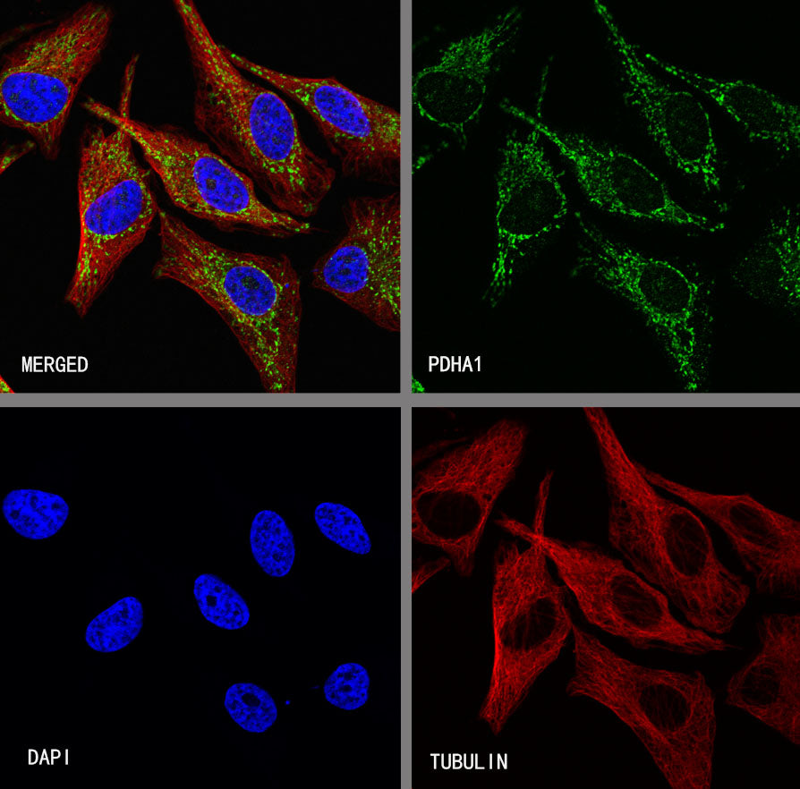
ICC shows positive staining in HeLa cells. Anti-PDHA1 antibody was used at 1/200 dilution (Green) and incubated overnight at 4°C. Goat polyclonal Antibody to Rabbit IgG - H&L (Alexa Fluor® 488) was used as secondary antibody at 1/1000 dilution. The cells were fixed with 4% PFA and permeabilized with 0.1% PBS-Triton X-100. Nuclei were counterstained with DAPI (Blue). Counterstain with tubulin (Red).