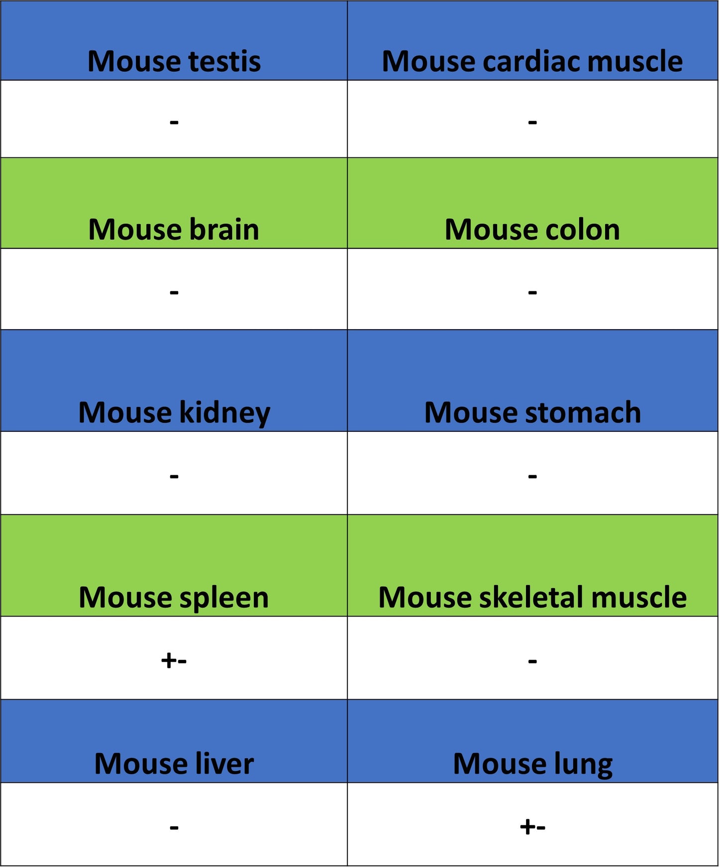
IHC shows positive staining in paraffin-embedded mouse lung. Anti-PD-L1 antibody was used at 1/250 dilution, followed by a HRP Polymer for Rabbit IgG (ready to use). Counterstained with hematoxylin. Heat mediated antigen retrieval with Tris/EDTA buffer pH9.0 was performed before commencing with IHC staining protocol.
PD-L1 Recombinant Rabbit mAb (S-5311)
PD-L1 Recombinant Rabbit mAb (S-5311)
Price:
Regular price
$100 USD
Regular price
Sale price
$100 USD
Unit price
per
For shipping services or bulk orders, you may request a quotation.
Secure checkout with

View full details
Product Details
Product Details
Product Specification
| Host | Rabbit |
| Antigen | CD274 |
| Synonyms | Programmed cell death 1 ligand 1; PD-L1; PDCD1 ligand 1; Programmed death ligand 1; hPD-L1; B7 homolog 1 (B7-H1); B7H1; PDCD1L1; PDCD1LG1; PDL1 |
| Location | Cell membrane |
| Accession | Q9NZQ7 |
| Clone Number | S-5311 |
| Antibody Type | Recombinant mAb |
| Isotype | IgG |
| Application | IHC-P |
| Reactivity | Ms |
| Purification | Protein A |
| Concentration | 0.5 mg/ml |
| Conjugation | Unconjugated |
| Physical Appearance | Liquid |
| Storage Buffer | PBS, 40% Glycerol, 0.05% BSA, 0.02% sodium azide |
| Stability & Storage | 12 months from date of receipt / reconstitution, -20 °C as supplied |
Dilution
| application | dilution | species |
| IHC-P | 1:250-1:500 | Ms |
Background
PD-L1 (Programmed Death-Ligand 1), is a ~33 kDa type I transmembrane glycoprotein of the immunoglobulin superfamily that functions as the key ligand for the inhibitory receptor PD-1 on T cells; upon IFN-γ–driven up-regulation on dendritic cells, macrophages, normal epithelia and many tumors, its engagement of PD-1 delivers a “stop” signal that blocks T-cell receptor signaling, suppressing proliferation, cytokine production and cytotoxic activity, thereby maintaining peripheral tolerance and preventing autoimmunity, but is exploited by cancers to evade immune destruction, making the PD-1/PD-L1 axis a central target for modern checkpoint-inhibitor immunotherapies.
Picture
Picture
Immunohistochemistry
IHC shows positive staining in paraffin-embedded mouse thymus. Anti-PD-L1 antibody was used at 1/250 dilution, followed by a HRP Polymer for Rabbit IgG (ready to use). Counterstained with hematoxylin. Heat mediated antigen retrieval with Tris/EDTA buffer pH9.0 was performed before commencing with IHC staining protocol.