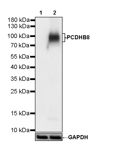
WB result of PCDHB8 Recombinant Rabbit mAb
Primary antibody: PCDHB8 Recombinant Rabbit mAb at 1/1000 dilution
Lane 1: untreated 293F whole cell lysate 20 µg
Lane 2: 293F transfected with PCDHB8-myc fusion protein whole cell lysate 20 µg
Secondary antibody: Goat Anti-Rabbit IgG (H+L), HRP conjugated at 1/10000 dilution
Predicted MW: 87 kDa
Observed MW: 87-100 kDa
PCDHB8 Recombinant Rabbit mAb (S-4950-10)
PCDHB8 Recombinant Rabbit mAb (S-4950-10)
Price:
Regular price
$100 USD
Regular price
Sale price
$100 USD
Unit price
per
For shipping services or bulk orders, you may request a quotation.
Secure checkout with

View full details
Product Details
Product Details
Product Specification
| Host | Rabbit |
| Antigen | PCDHB8 |
| Synonyms | Protocadherin beta-8; PCDH3I |
| Immunogen | Synthetic Peptide |
| Location | Cell membrane |
| Accession | Q9UN66 |
| Clone Number | S-4950-10 |
| Antibody Type | Recombinant mAb |
| Isotype | IgG |
| Application | WB |
| Reactivity | Hu |
| Purification | Protein A |
| Concentration | 0.5 mg/ml |
| Conjugation | Unconjugated |
| Physical Appearance | Liquid |
| Storage Buffer | PBS, 40% Glycerol, 0.05% BSA, 0.02% sodium azide |
| Stability & Storage | 12 months from date of receipt / reconstitution, -20 °C as supplied |
Dilution
| application | dilution | species |
| WB | 1:1000 | Hu |
Background
PCDHB8 (Protocadherin beta-8) is a calcium-dependent cell adhesion protein belonging to the clustered protocadherin beta subfamily within the non-classical cadherin superfamily. It is a single-pass type I membrane protein, with the human version comprising approximately 801 amino acids and having a theoretical molecular weight of about 87.6 kDa. Structurally, its extracellular region contains six cadherin repeat domains, each capable of binding three calcium ions. Among these, the Cadherin 1 through Cadherin 4 domains mediate homophilic trans dimerization with identical molecules on adjacent cell surfaces in a "head-to-tail" manner—Cadherin 1 of one molecule binds Cadherin 4 of the counterpart, and Cadherin 2 binds Cadherin 3—whereas the Cadherin 6 domain mediates promiscuous cis interactions with other protocadherins on the same cell membrane. Based on this molecular architecture, PCDHB8 is thought to participate in self/non-self discrimination between cells and thereby play a key role in the establishment and maintenance of specific neuronal connections in the central nervous system. At present, specific functional studies of this protein remain relatively limited, and its exact physiological role and significance in disease await further elucidation.
Picture
Picture
Western Blot