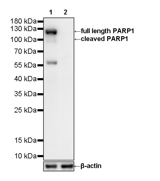
WB result of PARP1-Knockout HeLa Cell Lysate
Primary antibody: PARP1 Recombinant Rabbit mAb (S0B0650) at 1/1000 dilution
Lane 1: Wild-type HeLa whole cell lysate 20 ug
Lane 2: PARP1-Knockout HeLa whole Cell lysate 20 ug
Secondary antibody: Goat Anti-Rabbit IgG (H+L), HRP conjugated at 1/10000 dilution
Predicted MW: 113 kDa
Observed MW: 120 kDa
PARP1-Knockout HeLa Cell Lysate
PARP1-Knockout HeLa Cell Lysate
Price:
Regular price
$665 USD
Regular price
Sale price
$665 USD
Unit price
per
For shipping services or bulk orders, you may request a quotation.
Secure checkout with

View full details
Product Details
Product Details
Product Specification
| Host | Human |
| Mutations | Mutation in exon4 |
| Application | WB |
| Concentration | 2 mg/ml |
| Physical Appearance | Liquid |
| Storage Buffer | Supplied in SDS Sample Buffer: 50 mM Tris-HCl (pH 6.8 at 25°C), 2% w/v SDS, 10% glycerol, 1% v/v β-Mercaptoethanol, 0.01% w/v bromophenol blue or phenol red. |
| Stability & Storage | -20 °C for 1 months under sterile conditions; |
Background
PARP1 (poly [ADP-ribose] polymerase 1) is a highly abundant nuclear enzyme that plays a central role in the detection and repair of DNA damage. Upon sensing DNA single-strand breaks and other genomic lesions, PARP1 uses NAD+ as a substrate to synthesize poly (ADP-ribose) chains on itself and multiple target proteins, a process known as PARylation. This modification recruits and regulates DNA repair factors, promotes chromatin remodeling, and facilitates several DNA damage response pathways, particularly base excision repair and single-strand break repair. PARP1 is also involved in the regulation of transcription, replication stress responses, chromatin organization, and cell-death signaling. During apoptosis, PARP1 is cleaved by activated caspase-3 and caspase-7 into characteristic approximately 89 kDa and 24 kDa fragments; therefore, detection of cleaved PARP1 is widely used as a biochemical marker of caspase-dependent apoptosis. PARP1 antibodies are commonly used in Western blotting, immunohistochemistry, immunofluorescence, and flow cytometry to assess PARP1 expression, DNA damage responses, and apoptotic signaling. PARP1 knockout (KO) may impair efficient repair of DNA single-strand breaks, alter cellular responses to replication stress and genotoxic agents, and increase dependence on compensatory DNA repair pathways; however, the resulting phenotype can vary substantially with cell type, genetic background, and the nature of the DNA-damaging stimulus.
Picture
Picture
Western Blot