WB result of PAPSS1 Recombinant Rabbit mAb
Primary antibody: PAPSS1 Recombinant Rabbit mAb at 1/1000 dilution
Lane 1: THP-1 whole cell lysate 20 µg
Lane 2: HeLa whole cell lysate 20 µg
Lane 3: SK-OV-3 whole cell lysate 20 µg
Lane 4: Hep G2 whole cell lysate 20 µg
Lane 5: A673 whole cell lysate 20 µg
Lane 6: A549 whole cell lysate 20 µg
Lane 7: Ramos whole cell lysate 20 µg
Low expression control: THP-1 whole cell lysate
Secondary antibody: Goat Anti-Rabbit IgG (H+L), HRP conjugated at 1/10000 dilution
Predicted MW: 70 kDa
Observed MW: 70 kDa
PAPSS1 Recombinant Rabbit mAb (S-3516-23)
PAPSS1 Recombinant Rabbit mAb (S-3516-23)
Price:
Regular price
$100 USD
Regular price
Sale price
$100 USD
Unit price
per
For shipping services or bulk orders, you may request a quotation.
Secure checkout with

View full details
Product Details
Product Details
Product Specification
| Host | Rabbit |
| Antigen | PAPSS1 |
| Synonyms | Bifunctional 3'-phosphoadenosine 5'-phosphosulfate synthase 1; PAPS synthase 1; PAPSS 1Alternative nameSulfurylase kinase 1 (SK 1; SK1); ATPSK1; PAPSS |
| Immunogen | Recombinant Protein |
| Location | Nucleus |
| Accession | O43252 |
| Clone Number | S-3516-23 |
| Antibody Type | Recombinant mAb |
| Isotype | IgG |
| Application | WB, IHC-P |
| Reactivity | Hu, Ms, Rt |
| Positive Sample | HeLa, SK-OV-3, HepG2, A673, A594, Ramos, NIH/3T3, mouse testis, mouse colon, C6, rat testis, rat colon, COS7 |
| Purification | Protein A |
| Concentration | 0.5 mg/ml |
| Conjugation | Unconjugated |
| Physical Appearance | Liquid |
| Storage Buffer | PBS, 40% Glycerol, 0.05% BSA, 0.02% sodium azide |
| Stability & Storage | 12 months from date of receipt / reconstitution, -20 °C as supplied |
Dilution
| application | dilution | species |
| WB | 1:1000-1:5000 | Hu, Ms, Rt, Mk |
| IHC-P | 1:500-1:2000 | Hu, Ms, Rt |
Background
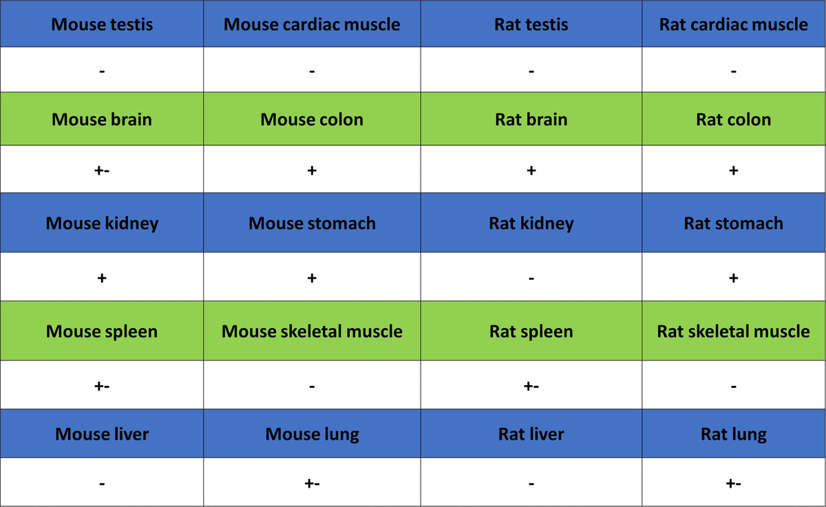
PAPSS1 (3'-phosphoadenosine-5'-phosphosulfate synthase 1) is a bifunctional enzyme and the only key protein in living organisms capable of catalyzing sulfate activation. Its core function is to catalyze, through its N-terminal APS kinase domain and C-terminal ATP sulfurylase domain, the two-step generation of the activated sulfate donor PAPS (3'-phosphoadenosine-5'-phosphosulfate) using ATP as an energy source, thereby providing the essential sulfate group for all downstream sulfotransferases. Structurally, PAPSS1 exists as a homodimer, and its unique nuclear localization signal directs it predominantly to the cell nucleus—a striking contrast to the mainly cytoplasmic localization of its family member PAPSS2—suggesting the existence of an independent sulfation pathway within the nucleus, with the C-terminal domain also exerting regulatory effects on the N-terminal kinase activity. Functionally, PAPSS1 is widely and highly expressed in most tissues including the brain (except the liver), and the PAPS it generates is critical for synthesizing sulfated L-selectin ligands on endothelial cell surfaces that mediate lymphocyte homing. Furthermore, recent studies have unveiled novel roles of PAPSS1 under pathological conditions: for example, in ovarian cancer, its high expression is closely associated with platinum-based chemotherapy resistance, and inhibiting PAPSS1 can significantly restore cancer cell sensitivity to cisplatin by enhancing estrogen receptor α (ERα) signaling, making it a highly promising target for chemosensitization.
Picture
Picture
Western Blot
WB result of PAPSS1 Recombinant Rabbit mAb
Primary antibody: PAPSS1 Recombinant Rabbit mAb at 1/1000 dilution
Lane 1: NIH/3T3 whole cell lysate 20 µg
Lane 2: mouse testis lysate 20 µg
Lane 3: mouse colon lysate 20 µg
Secondary antibody: Goat Anti-Rabbit IgG (H+L), HRP conjugated at 1/10000 dilution
Predicted MW: 70 kDa
Observed MW: 70 kDa
WB result of PAPSS1 Recombinant Rabbit mAb
Primary antibody: PAPSS1 Recombinant Rabbit mAb at 1/1000 dilution
Lane 1: C6 whole cell lysate 20 µg
Lane 2: rat testis lysate 20 µg
Lane 3: rat colon lysate 20 µg
Secondary antibody: Goat Anti-Rabbit IgG (H+L), HRP conjugated at 1/10000 dilution
Predicted MW: 70 kDa
Observed MW: 70 kDa
WB result of PAPSS1 Recombinant Rabbit mAb
Primary antibody: PAPSS1 Recombinant Rabbit mAb at 1/1000 dilution
Lane 1: COS7 whole cell lysate 20 µg
Secondary antibody: Goat Anti-Rabbit IgG (H+L), HRP conjugated at 1/10000 dilution
Predicted MW: 70 kDa
Observed MW: 70 kDa
Immunohistochemistry
IHC shows positive staining in paraffin-embedded human colon. Anti-PAPSS1 antibody was used at 1/500 dilution, followed by a HRP Polymer for Rabbit IgG (ready to use). Counterstained with hematoxylin. Heat mediated antigen retrieval with Tris/EDTA buffer pH9.0 was performed before commencing with IHC staining protocol.
IHC shows positive staining in paraffin-embedded human testis. Anti-PAPSS1 antibody was used at 1/500 dilution, followed by a HRP Polymer for Rabbit IgG (ready to use). Counterstained with hematoxylin. Heat mediated antigen retrieval with Tris/EDTA buffer pH9.0 was performed before commencing with IHC staining protocol.
IHC shows positive staining in paraffin-embedded human cervical squamous cell carcinoma. Anti-PAPSS1 antibody was used at 1/2000 dilution, followed by a HRP Polymer for Rabbit IgG (ready to use). Counterstained with hematoxylin. Heat mediated antigen retrieval with Tris/EDTA buffer pH9.0 was performed before commencing with IHC staining protocol.
IHC shows positive staining in paraffin-embedded human lung cancer. Anti-PAPSS1 antibody was used at 1/2000 dilution, followed by a HRP Polymer for Rabbit IgG (ready to use). Counterstained with hematoxylin. Heat mediated antigen retrieval with Tris/EDTA buffer pH9.0 was performed before commencing with IHC staining protocol.
IHC shows positive staining in paraffin-embedded human ovarian cancer. Anti-PAPSS1 antibody was used at 1/2000 dilution, followed by a HRP Polymer for Rabbit IgG (ready to use). Counterstained with hematoxylin. Heat mediated antigen retrieval with Tris/EDTA buffer pH9.0 was performed before commencing with IHC staining protocol.
IHC shows positive staining in paraffin-embedded mouse colon. Anti-PAPSS1 antibody was used at 1/2000 dilution, followed by a HRP Polymer for Rabbit IgG (ready to use). Counterstained with hematoxylin. Heat mediated antigen retrieval with Tris/EDTA buffer pH9.0 was performed before commencing with IHC staining protocol.
IHC shows positive staining in paraffin-embedded rat colon. Anti-PAPSS1 antibody was used at 1/2000 dilution, followed by a HRP Polymer for Rabbit IgG (ready to use). Counterstained with hematoxylin. Heat mediated antigen retrieval with Tris/EDTA buffer pH9.0 was performed before commencing with IHC staining protocol.