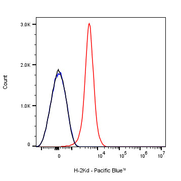
Flow cytometric analysis of Mouse H-2Kd expression on BALB/c mouse splenocytes. BALB/c mouse splenocytes were stained with Pacific Blue™ Mouse IgG2a, κ Isotype Control (Black line histogram) or SDT Pacific Blue™ Mouse Anti-Mouse H-2Kd Antibody (Red line histogram) at 5μl/test, cells without incubation with primary antibody and secondary antibody (Blue line histogram) was used as unlabelled control. Total viable cells, as determined by Fixable Viability Dye 662 (S0D0016), were used for analysis. Flow cytometry and data analysis were performed using Agilent NovoCyte Quanteon and FlowJo™ software.
Pacific Blue Mouse Anti-Mouse H-2Kd Antibody (SF1-1.1)
Pacific Blue Mouse Anti-Mouse H-2Kd Antibody (SF1-1.1)
Price:
Regular price
$97 USD
Regular price
Sale price
$97 USD
Unit price
per
For shipping services or bulk orders, you may request a quotation.
Secure checkout with

View full details
Product Details
Product Details
Product Specification
| Host | Mouse |
| Antigen | H-2Kd |
| Synonyms | H-2 class I histocompatibility antigen, K-D alpha chain; H-2K(D); H2-K; H2-K1 |
| Location | Membrane |
| Accession | P01902 |
| Clone Number | SF1-1.1 |
| Antibody Type | Mouse mAb |
| Isotype | IgG2a,k |
| Application | FCM |
| Reactivity | Ms |
| Positive Sample | Balb/c mouse splenocytes |
| Purification | Protein A |
| Concentration | 0.2 mg/ml |
| Conjugation | Pacific Blue |
| Physical Appearance | Liquid |
| Storage Buffer | PBS, 1% BSA, 0.3% Proclin 300 |
| Stability & Storage | 12 months from date of receipt / reconstitution, 2 to 8 °C as supplied |
Dilution
| application | dilution | species |
| FCM | 5μl per million cells in 100μl volume | Ms |
Background
H-2Kd is a murine MHC class I molecule expressed by BALB/c mice that presents endogenous 9-mer peptides—anchored by Tyr at position 2 and a hydrophobic residue at position 9—to CD8⁺ cytotoxic T cells, thereby initiating adaptive immune responses; its 2.6-Å crystal structure reveals a peptide-binding groove with five deep pockets (A–F) that impose an allele-specific motif, and it pairs non-covalently with β2-microglobulin for stability.
Picture
Picture
FC