WB result of PABPN1 Rabbit Polyclonal Antibody
Primary antibody: PABPN1 Rabbit Polyclonal Antibody at 1/1000 dilution
Lane 1: 293T whole cell lysate 20 μg
Lane 2: MCF7 whole cell lysate 20 μg
Lane 3: HeLa whole cell lysate 20 μg
Lane 4: K-562 whole cell lysate 20 μg
Secondary antibody: Goat Anti-Rabbit IgG (H+L), HRP conjugated at 1/10000 dilution
Predicted MW: 33 kDa
Observed MW: 50 kDa
PABPN1 Rabbit Polyclonal Antibody
PABPN1 Rabbit Polyclonal Antibody
Price:
Regular price
$100 USD
Regular price
Sale price
$100 USD
Unit price
per
For shipping services or bulk orders, you may request a quotation.
Secure checkout with

View full details
Product Details
Product Details
Product Specification
| Host | Rabbit |
| Antigen | PABPN1 |
| Immunogen | Synthetic Peptide |
| Location | Cytoplasm, Nucleus |
| Accession | Q86U42 |
| Antibody Type | Polyclonal antibody |
| Isotype | IgG |
| Application | WB, IHC-P, ICC, IF |
| Reactivity | Hu, Ms, Rt, Mk |
| Positive Sample | 293T, MCF7, HeLa, K-562, RAW264.7, mouse spleen, PC-12, rat spleen, COS-7 |
| Purification | Protein A |
| Concentration | 2 mg/ml |
| Conjugation | Unconjugated |
| Physical Appearance | Liquid |
| Storage Buffer | PBS, 40% Glycerol, 0.05% BSA, 0.02% sodium azide |
| Stability & Storage | 12 months from date of receipt / reconstitution, -20 °C as supplied |
Dilution
| application | dilution | species |
| WB | 1:1000-1:2000 | Hu, Ms, Rt, Mk |
| IHC-P | 1:800-1:4000 | Hu, Ms, Rt |
| ICC | 1:2000 | Hu, Ms |
| IF | 1:4000 | Hu |
Background
PABPN1 (poly(A)-binding protein nuclear 1) is the only poly(A)-binding protein found in the nucleus of mammals, encoded by the PABPN1 gene located on human chromosome 14q11.2, with a molecular weight of approximately 32.7 kDa and containing 306 amino acids. Its structure consists of an N-terminal alanine-rich intrinsically disordered region (IDR), a central coiled-coil domain (CCD), and a C-terminal RNA recognition motif (RRM), in which the N-terminal IDR serves as a "gatekeeper" for protein interactions, while the N-terminal region of the CCD determines protein stability and the C-terminal region expands the interactome. Functionally, PABPN1 binds the nascent poly(A) tail with high affinity and regulates the gradual extension of the poly(A) tail from approximately 10 adenosines to about 250 nucleotides, thereby controlling mRNA 3' end processing and alternative polyadenylation (APA). Loss of PABPN1 function leads to genome-wide APA shifts, affecting mRNA nuclear export and translation. Clinically, abnormal expansion of the GCG trinucleotide repeat at the 5' end of the PABPN1 coding region (expanding from the normal 6 copies to 8-13 copies) causes autosomal dominant oculopharyngeal muscular dystrophy (OPMD), the pathological mechanism of which involves alanine-expanded PABPN1 forming insoluble intranuclear aggregates and reducing functional protein levels. In addition, PABPN1 is abnormally expressed in tumors such as bladder cancer and participates in hematopoietic stem cell maintenance.
Picture
Picture
Western Blot
WB result of PABPN1 Rabbit Polyclonal Antibody
Primary antibody: PABPN1 Rabbit Polyclonal Antibody at 1/1000 dilution
Lane 1: Raw 264.7 whole cell lysate 20 μg
Lane 2: mouse spleen lysate 20 μg
Secondary antibody: Goat Anti-Rabbit IgG (H+L), HRP conjugated at 1/10000 dilution
Predicted MW: 33 kDa
Observed MW: 50 kDa
WB result of PABPN1 Rabbit Polyclonal Antibody
Primary antibody: PABPN1 Rabbit Polyclonal Antibody at 1/1000 dilution
Lane 1: PC-12 whole cell lysate 20 μg
Lane 2: rat spleen lysate 20 μg
Secondary antibody: Goat Anti-Rabbit IgG (H+L), HRP conjugated at 1/10000 dilution
Predicted MW: 33 kDa
Observed MW: 50 kDa
WB result of PABPN1 Rabbit Polyclonal Antibody
Primary antibody: PABPN1 Rabbit Polyclonal Antibody at 1/1000 dilution
Lane 1: COS-7 whole cell lysate 20 μg
Secondary antibody: Goat Anti-Rabbit IgG (H+L), HRP conjugated at 1/10000 dilution
Predicted MW: 33 kDa
Observed MW: 50 kDa
Immunohistochemistry
IHC shows positive staining in paraffin-embedded human kidney. Anti-PABPN1 antibody was used at 1/4000 dilution, followed by a HRP Polymer for Rabbit IgG (ready to use). Counterstained with hematoxylin. Heat mediated antigen retrieval with Tris/EDTA buffer pH9.0 was performed before commencing with IHC staining protocol.
IHC shows positive staining in paraffin-embedded human lung cancer. Anti-PABPN1 antibody was used at 1/4000 dilution, followed by a HRP Polymer for Rabbit IgG (ready to use). Counterstained with hematoxylin. Heat mediated antigen retrieval with Tris/EDTA buffer pH9.0 was performed before commencing with IHC staining protocol.
IHC shows positive staining in paraffin-embedded mouse testis. Anti-PABPN1 antibody was used at 1/4000 dilution, followed by a HRP Polymer for Rabbit IgG (ready to use). Counterstained with hematoxylin. Heat mediated antigen retrieval with Tris/EDTA buffer pH9.0 was performed before commencing with IHC staining protocol.
IHC shows positive staining in paraffin-embedded rat testis. Anti-PABPN1 antibody was used at 1/4000 dilution, followed by a HRP Polymer for Rabbit IgG (ready to use). Counterstained with hematoxylin. Heat mediated antigen retrieval with Tris/EDTA buffer pH9.0 was performed before commencing with IHC staining protocol.
Immunocytochemistry
ICC shows positive staining in 293T cells. Anti-PABPN1 antibody was used at 1/2000 dilution (Green) and incubated overnight at 4°C. Goat polyclonal Antibody to Rabbit IgG - H&L (Alexa Fluor® 488) was used as secondary antibody at 1/1000 dilution. The cells were fixed with 4% PFA and permeabilized with 0.1% PBS-Triton X-100. Nuclei were counterstained with DAPI (Blue). Counterstain with tubulin (Red).
ICC shows positive staining in RAW 264.7 cells. Anti-PABPN1 antibody was used at 1/2000 dilution (Green) and incubated overnight at 4°C. Goat polyclonal Antibody to Rabbit IgG - H&L (Alexa Fluor® 488) was used as secondary antibody at 1/1000 dilution. The cells were fixed with 4% PFA and permeabilized with 0.1% PBS-Triton X-100. Nuclei were counterstained with DAPI (Blue). Counterstain with tubulin (Red).
Immunofluorescence
IF shows positive staining in paraffin-embedded human colon. Anti-PABPN1 antibody was used at 1/4000 dilution (Green) and incubated overnight at 4°C. Goat polyclonal Antibody to Rabbit IgG - H&L (Alexa Fluor® 488) was used as secondary antibody at 1/1000 dilution. Counterstained with DAPI (Blue). Heat mediated antigen retrieval with EDTA buffer pH9.0 was performed before commencing with IF staining protocol.
IF shows positive staining in paraffin-embedded human testis. Anti-PABPN1 antibody was used at 1/4000 dilution (Green) and incubated overnight at 4°C. Goat polyclonal Antibody to Rabbit IgG - H&L (Alexa Fluor® 488) was used as secondary antibody at 1/1000 dilution. Counterstained with DAPI (Blue). Heat mediated antigen retrieval with EDTA buffer pH9.0 was performed before commencing with IF staining protocol.
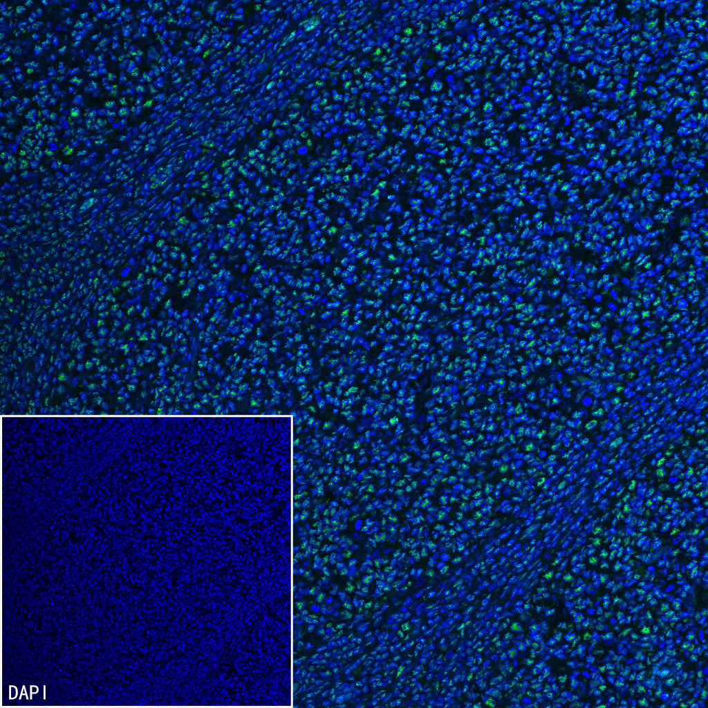
IF shows positive staining in paraffin-embedded human tonsil. Anti-PABPN1 antibody was used at 1/4000 dilution (Green) and incubated overnight at 4°C. Goat polyclonal Antibody to Rabbit IgG - H&L (Alexa Fluor® 488) was used as secondary antibody at 1/1000 dilution. Counterstained with DAPI (Blue). Heat mediated antigen retrieval with EDTA buffer pH9.0 was performed before commencing with IF staining protocol.