WB result of p57 Kip2 Recombinant Rabbit mAb
Primary antibody: p57 Kip2 Recombinant Rabbit mAb at 1/1000 dilution
Lane 1: untreated HeLa whole cell lysate 20 μg
Lane 2: HeLa treated with 50nM Dexamethasone for 16 hours whole cell lysate 20 μg
Secondary antibody: Goat Anti-Rabbit IgG (H+L), HRP conjugated at 1/10000 dilution
Predicted MW: 32 kDa
Observed MW: 57 kDa
p57 Kip2 Recombinant Rabbit mAb (S-6119)
p57 Kip2 Recombinant Rabbit mAb (S-6119)
Price:
Regular price
$100 USD
Regular price
Sale price
$100 USD
Unit price
per
For shipping services or bulk orders, you may request a quotation.
Secure checkout with

View full details
Product Details
Product Details
Product Specification
| Host | Rabbit |
| Antigen | p57 Kip2 |
| Location | Nucleus |
| Accession | P49918 |
| Clone Number | S-6119 |
| Antibody Type | Recombinant mAb |
| Isotype | IgG |
| Application | WB, IHC-P |
| Reactivity | Hu |
| Purification | Protein A |
| Concentration | 0.5 mg/ml |
| Conjugation | Unconjugated |
| Physical Appearance | Liquid |
| Storage Buffer | PBS, 40% Glycerol, 0.05% BSA, 0.02% sodium azide |
| Stability & Storage | 12 months from date of receipt / reconstitution, -20 °C as supplied |
Dilution
| application | dilution | species |
| WB | 1:1000-1:2000 | Hu |
| IHC-P | 1:2000 | Hu |
Background
p57 Kip2 (also known as CDKN1C) is a member of the Cip/Kip family encoded by the paternally imprinted gene CDKN1C, and is the only member of this family that is essential for embryonic development. Human CDKN1C is located on chromosome 11p15.5 and is expressed only from the maternal allele, with its transcription regulated by the ICR2 imprinting control region and the KCNQ1OT1 non-coding RNA. p57 Kip2 is a potent, tightly binding inhibitor of G1-phase cyclin/CDK complexes (such as cyclin E-CDK2 and cyclin D2-CDK4), and its overexpression can cause complete cell cycle arrest in G1 phase. Its structure consists of an N-terminal CDK inhibitory domain, a proline-rich domain, an acidic repeat region, and a C-terminal nuclear localization domain, with nuclear localization depending on a classical bipartite nuclear localization signal formed by two positively charged clusters. Functionally, p57 Kip2 is highly expressed in terminally differentiated tissues such as skeletal muscle, heart, brain, and lung, participating in cell cycle exit and differentiation regulation. Loss of its function is associated with Beckwith-Wiedemann syndrome (overgrowth), while missense mutations in the PCNA-binding domain cause IMAGe syndrome (growth restriction). In addition, p57 Kip2 expression is downregulated in various cancers, but mutations in the gene itself are rare, suggesting that its tumor suppressor function is mainly inactivated through epigenetic silencing.
Picture
Picture
Western Blot
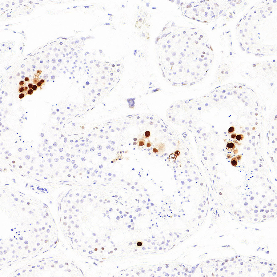
Immunohistochemistry
IHC shows positive staining in paraffin-embedded human kidney. Anti-p57 Kip2 antibody was used at 1/2000 dilution, followed by a HRP Polymer for Rabbit IgG (ready to use). Counterstained with hematoxylin. Heat mediated antigen retrieval with Tris/EDTA buffer pH9.0 was performed before commencing with IHC staining protocol.
IHC shows positive staining in paraffin-embedded human placenta. Anti-p57 Kip2 antibody was used at 1/2000 dilution, followed by a HRP Polymer for Rabbit IgG (ready to use). Counterstained with hematoxylin. Heat mediated antigen retrieval with Tris/EDTA buffer pH9.0 was performed before commencing with IHC staining protocol.
IHC shows positive staining in paraffin-embedded human testis. Anti-p57 Kip2 antibody was used at 1/2000 dilution, followed by a HRP Polymer for Rabbit IgG (ready to use). Counterstained with hematoxylin. Heat mediated antigen retrieval with Tris/EDTA buffer pH9.0 was performed before commencing with IHC staining protocol.