WB result of NSG2 Recombinant Rabbit mAb
Primary antibody: NSG2 Recombinant Rabbit mAb at 1/1000 dilution
Lane 1: Raji whole cell lysate 20 µg
Lane 2: SH-SY5Y whole cell lysate 20 µg
Lane 3: Neuro-2a whole cell lysate 20 µg
Negative control: Raji whole cell lysate
Secondary antibody: Goat Anti-Rabbit IgG (H+L), HRP conjugated at 1/10000 dilution
Predicted MW: 19 kDa
Observed MW: 17, 20 kDa
NSG2 Recombinant Rabbit mAb (S-3789-136)
NSG2 Recombinant Rabbit mAb (S-3789-136)
Price:
Regular price
$100 USD
Regular price
Sale price
$100 USD
Unit price
per
For shipping services or bulk orders, you may request a quotation.
Secure checkout with

View full details
Product Details
Product Details
Product Specification
| Host | Rabbit |
| Antigen | NSG2 |
| Synonyms | Neuronal vesicle trafficking-associated protein 2; Neuron-specific protein family member 2Imported; Protein p19 (Hmp19); CALY3 |
| Immunogen | Synthetic Peptide |
| Location | Lysosome, Endosome |
| Accession | Q9Y328 |
| Clone Number | S-3789-136 |
| Antibody Type | Recombinant mAb |
| Isotype | IgG |
| Application | WB, ICC |
| Reactivity | Hu, Ms, Rt |
| Positive Sample | SH-SY5Y, Neuro-2a, mouse brain, rat brain |
| Purification | Protein A |
| Concentration | 2 mg/ml |
| Conjugation | Unconjugated |
| Physical Appearance | Liquid |
| Storage Buffer | PBS, 40% Glycerol, 0.05% BSA, 0.02% sodium azide |
| Stability & Storage | 12 months from date of receipt / reconstitution, -20 °C as supplied |
Dilution
| application | dilution | species |
| WB | 1:1000 | Hu, Ms, Rt |
| ICC | 1:500 | Hu |
Background
NSG2, also known as neuronal vesicle trafficking-associated protein 2, is a membrane protein encoded by the NSG2 gene that is predominantly and specifically expressed in brain tissue. It is localized to the Golgi apparatus and multivesicular bodies and participates in clathrin-mediated intracellular trafficking processes. This protein consists of 171 amino acids with a molecular weight of approximately 19 kDa, belongs to the NSG family, and is also referred to as p19 or HMP19. As an AMPA receptor-binding protein, NSG2 regulates the recycling trafficking and surface levels of postsynaptic AMPA receptors, thereby playing an important regulatory role in excitatory neurotransmission and synaptic plasticity. Notably, gene knockout studies in mouse models have shown that loss of NSG2 does not lead to behavioral deficits but instead selectively enhances various associative learning and memory abilities, including spatial learning, reversal learning, and trace fear conditioning, suggesting that NSG2 may be a potential target for cognitive enhancement.
Picture
Picture
Western Blot
WB result of NSG2 Recombinant Rabbit mAb
Primary antibody: NSG2 Recombinant Rabbit mAb at 1/1000 dilution
Lane 1: mouse brain lysate 20 µg
Secondary antibody: Goat Anti-Rabbit IgG (H+L), HRP conjugated at 1/10000 dilution
Predicted MW: 19 kDa
Observed MW: 17, 20 kDa
WB result of NSG2 Recombinant Rabbit mAb
Primary antibody: NSG2 Recombinant Rabbit mAb at 1/1000 dilution
Lane 1: rat brain lysate 20 µg
Secondary antibody: Goat Anti-Rabbit IgG (H+L), HRP conjugated at 1/10000 dilution
Predicted MW: 19 kDa
Observed MW: 17, 20 kDa
Immunocytochemistry
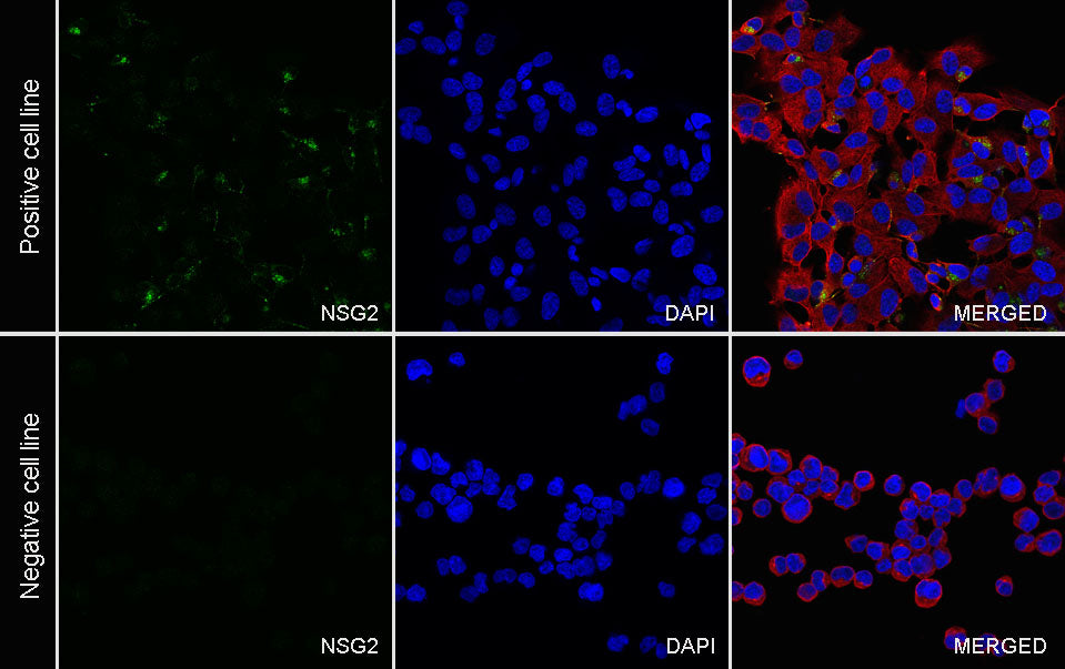
ICC shows positive staining in SH-SY5Y cells (top panel) and negative staining in Raji cells (below panel). Anti-NSG2 antibody was used at 1/500 dilution (Green) and incubated overnight at 4°C. Goat polyclonal Antibody to Rabbit IgG - H&L (Alexa Fluor® 488) was used as secondary antibod at 1/1000 dilution. The cells were fixed with 4% PFA and permeabilized with 0.1% PBS-Triton X-100. Nuclei were counterstained with DAPI (Blue). Counterstain with tubulin (Red).