WB result of Noxa Recombinant Rabbit mAb
Primary antibody: Noxa Recombinant Rabbit mAb at 1/1000 dilution
Lane 1: untreated Jurkat whole cell lysate 20 µg
Lane 2: Jurkat treated with 10 ng/ml TPA for 3 hours whole cell lysate 20 µg
Secondary antibody: Goat Anti-rabbit IgG (H+L), HRP conjugated at 1/10000 dilution
Predicted MW: 51 kDa
Observed MW: 12 kDa
Noxa Recombinant Rabbit mAb (S-5505)
Noxa Recombinant Rabbit mAb (S-5505)
Price:
Regular price
$100 USD
Regular price
Sale price
$100 USD
Unit price
per
For shipping services or bulk orders, you may request a quotation.
Secure checkout with

View full details
Product Details
Product Details
Product Specification
| Host | Rabbit |
| Antigen | Noxa |
| Synonyms | NADPH oxidase activator 1; Antigen NY-CO-31; NCF2-like protein; P67phox-like factor; p51-nox; P51NOX; NOXA1 |
| Location | Cytoplasm, Cell membrane |
| Accession | Q86UR1 |
| Clone Number | S-5505 |
| Antibody Type | Recombinant mAb |
| Isotype | IgG |
| Application | WB, IHC-P |
| Reactivity | Hu |
| Predicted Reactivity | Ms |
| Purification | Protein A |
| Concentration | 2 mg/ml |
| Conjugation | Unconjugated |
| Physical Appearance | Liquid |
| Storage Buffer | PBS, 40% Glycerol, 0.05% BSA, 0.02% sodium azide |
| Stability & Storage | 12 months from date of receipt / reconstitution, -20 °C as supplied |
Dilution
| application | dilution | species |
| WB | 1:1000-1:5000 | Hu |
| IHC-P | 1:1000 | Hu |
Background
Noxa, also known as PMA-induced protein 1 (PMAIP1), is a pro-apoptotic member of the Bcl-2 protein family that functions as a critical mediator of p53-dependent and -independent apoptosis by selectively neutralizing anti-apoptotic proteins Mcl-1 and A1/Bfl-1 through its BH3-only domain. Transcriptionally upregulated in response to diverse cellular stresses including DNA damage, hypoxia, proteasome inhibition, and oncogene activation, Noxa promotes mitochondrial outer membrane permeabilization (MOMP) not by directly activating Bax or Bak, but by displacing them from their inhibitory interactions with Mcl-1, thereby enabling cytochrome c release and caspase cascade initiation. Its expression is tightly regulated at both transcriptional and post-translational levels, and its functional specificity for Mcl-1 makes it a key determinant of cellular sensitivity to therapies targeting this anti-apoptotic protein, particularly in cancers where Mcl-1 overexpression confers resistance; consequently, Noxa serves as both a biomarker of stress-induced apoptotic priming and a potential therapeutic target for sensitizing tumor cells to chemotherapy and targeted agents.
Picture
Picture
Western Blot
Immunohistochemistry
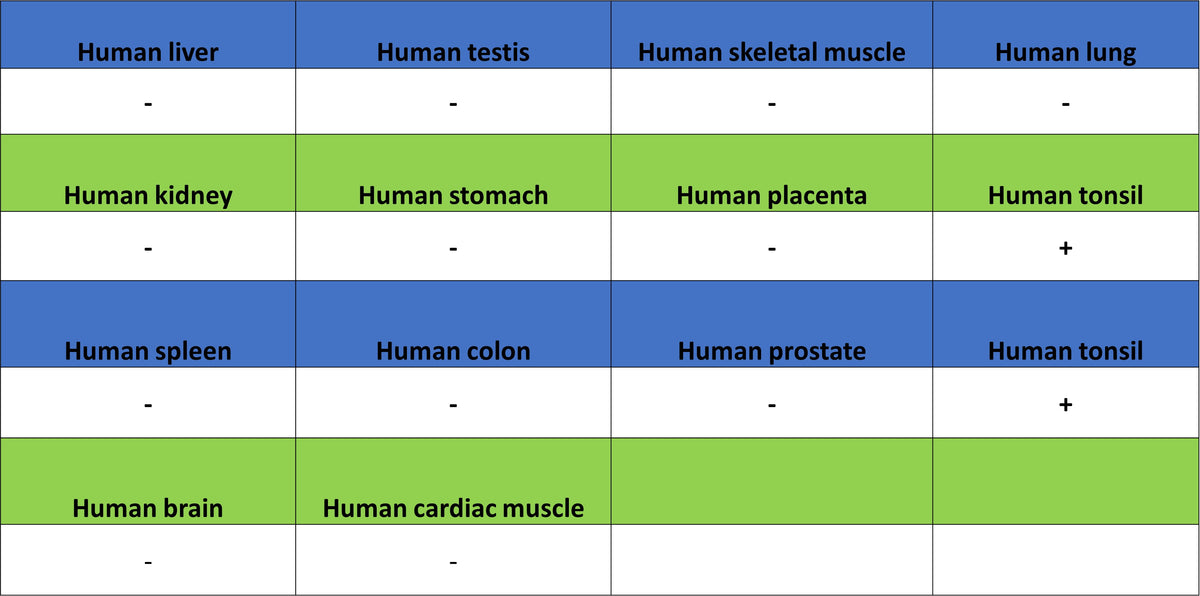
IHC shows positive staining in paraffin-embedded human tonsil. Anti-Noxa antibody was used at 1/1000 dilution, followed by a HRP Polymer for Rabbit IgG (ready to use). Counterstained with hematoxylin. Heat mediated antigen retrieval with Tris/EDTA buffer pH9.0 was performed before commencing with IHC staining protocol.
IHC shows positive staining in paraffin-embedded human lung cancer. Anti-Noxa antibody was used at 1/1000 dilution, followed by a HRP Polymer for Rabbit IgG (ready to use). Counterstained with hematoxylin. Heat mediated antigen retrieval with Tris/EDTA buffer pH9.0 was performed before commencing with IHC staining protocol.
IHC shows positive staining in paraffin-embedded human diffuse large B-cell lymphoma. Anti-Noxa antibody was used at 1/1000 dilution, followed by a HRP Polymer for Rabbit IgG (ready to use). Counterstained with hematoxylin. Heat mediated antigen retrieval with Tris/EDTA buffer pH9.0 was performed before commencing with IHC staining protocol.