WB result of NOL3/ARC Recombinant Rabbit mAb
Primary antibody: NOL3/ARC Recombinant Rabbit mAb at 1/1000 dilution
Lane 1: Daudi whole cell lysate 20 µg
Lane 2: RPMI-8226 whole cell lysate 20 µg
Lane 3: HCT-116 whole cell lysate 20 µg
Lane 4: MCF7 whole cell lysate 20 µg
Negative control: Daudi whloe cell lysate
Secondary antibody: Goat Anti-Rabbit IgG (H+L), HRP conjugated at 1/10000 dilution
Predicted MW: 23 kDa
Observed MW: 27 kDa
NOL3/ARC Recombinant Rabbit mAb (S-4660-38)
NOL3/ARC Recombinant Rabbit mAb (S-4660-38)
Price:
Regular price
$100 USD
Regular price
Sale price
$100 USD
Unit price
per
For shipping services or bulk orders, you may request a quotation.
Secure checkout with

View full details
Product Details
Product Details
Product Specification
| Host | Rabbit |
| Antigen | NOL3/ARC |
| Synonyms | Nucleolar protein 3; Apoptosis repressor with CARD; Muscle-enriched cytoplasmic protein (Myp); Nucleolar protein of 30 kDa (Nop30); NOP |
| Immunogen | Synthetic Peptide |
| Location | Cytoplasm, Nucleus |
| Accession | O60936 |
| Clone Number | S-4660-38 |
| Antibody Type | Recombinant mAb |
| Isotype | IgG |
| Application | WB |
| Reactivity | Hu, Ms, Rt |
| Positive Sample | RPMI-8226, HCT-116, mouse cardiac muscle, mouse skeletal muscle, rat skeletal muscle |
| Purification | Protein A |
| Concentration | 0.5 mg/ml |
| Conjugation | Unconjugated |
| Physical Appearance | Liquid |
| Storage Buffer | PBS, 40% Glycerol, 0.05% BSA, 0.02% sodium azide |
| Stability & Storage | 12 months from date of receipt / reconstitution, -20 °C as supplied |
Dilution
| application | dilution | species |
| WB | 1:1000-1:5000 | Hu, Ms, Rt |
Background
NOL3 (Nucleolar Protein 3), also known as ARC (Apoptosis Repressor with CARD Domain), is a multifunctional anti-apoptotic protein with dual subcellular localization. Through its N-terminal CARD domain, it can simultaneously block both extrinsic and intrinsic apoptotic signaling pathways through multiple mechanisms. On one hand, NOL3 interferes with the assembly of the death-inducing signaling complex by binding to FAS and FADD, or restricts the activation of Caspase-8 by binding to it, thereby inhibiting the extrinsic apoptotic pathway; on the other hand, it can directly bind and inactivate the pro-apoptotic protein BAX, preventing its translocation from the cytoplasm to the mitochondria, thereby inhibiting mitochondrial outer membrane permeabilization and cytochrome c release, effectively blocking the intrinsic apoptotic pathway. Furthermore, NOL3 can bind to p53 to inhibit its tetramerization and transcriptional function, and through phosphorylation modifications it regulates its binding to Caspase-8 to exert calcium-buffering effects, maintaining cell survival at multiple levels. In terms of tissue distribution, NOL3 is highly expressed in heart and skeletal muscle. Under pathological conditions, its protein level is rapidly degraded via the ubiquitin-proteasome pathway during myocardial ischemia-reperfusion, making it a critical regulatory node in myocardial injury; meanwhile, NOL3 is overexpressed in various epithelial-derived tumors (such as breast cancer and renal cancer), closely associated with chemoresistance and radioresistance; and its gene mutations have been confirmed to be associated with neurological disorders such as familial cortical myoclonus.
Picture
Picture
Western Blot
WB result of NOL3/ARC Recombinant Rabbit mAb
Primary antibody: NOL3/ARC Recombinant Rabbit mAb at 1/1000 dilution
Lane 1: mouse liver lysate 20 µg
Lane 2: mouse cardiac muscle lysate 20 µg
Lane 3: mouse skeletal muscle lysate 20 µg
Negative control: mouse liver lysate
Secondary antibody: Goat Anti-Rabbit IgG (H+L), HRP conjugated at 1/10000 dilution
Predicted MW: 23 kDa
Observed MW: 27 kDa
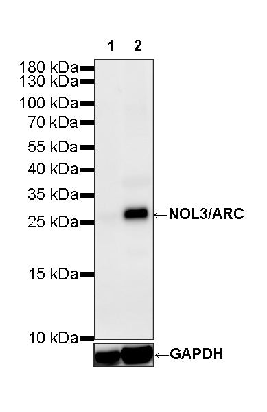
WB result of NOL3/ARC Recombinant Rabbit mAb
Primary antibody: NOL3/ARC Recombinant Rabbit mAb at 1/1000 dilution
Lane 1: rat liver lysate 20 µg
Lane 2: rat skeletal muscle lysate 20 µg
Negative control: rat liver lysate
Secondary antibody: Goat Anti-Rabbit IgG (H+L), HRP conjugated at 1/10000 dilution
Predicted MW: 23 kDa
Observed MW: 27 kDa