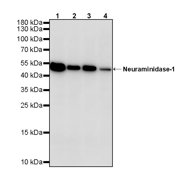
WB result of Neuraminidase-1 Recombinant Rabbit mAb
Primary antibody: Neuraminidase-1 Recombinant Rabbit mAb at 1/1000 dilution
Lane 1: unboiled HeLa whole cell lysate 20 µg
Lane 2: unboiled A549 whole cell lysate 20 µg
Lane 3: unboiled Hep G2 whole cell lysate 20 µg
Lane 4: unboiled A-375 whole cell lysate 20 µg
Secondary antibody: Goat Anti-rabbit IgG (H+L), HRP conjugated at 1/10000 dilution
Predicted MW: 45 kDa
Observed MW: 50 kDa
This blot was developed with high sensitivity substrate
Neuraminidase-1 Recombinant Rabbit mAb (S-4004-109)
Neuraminidase-1 Recombinant Rabbit mAb (S-4004-109)
Price:
Regular price
$100 USD
Regular price
Sale price
$100 USD
Unit price
per
For shipping services or bulk orders, you may request a quotation.
Secure checkout with

View full details
Product Details
Product Details
Product Specification
| Host | Rabbit |
| Antigen | Neuraminidase-1 |
| Immunogen | Synthetic Peptide |
| Location | Lysosome, Cell membrane |
| Accession | Q99519 |
| Clone Number | S-4004-109 |
| Antibody Type | Recombinant mAb |
| Isotype | IgG |
| Application | WB |
| Reactivity | Hu |
| Positive Sample | HeLa, A549, Hep G2, A-375 |
| Purification | Protein A |
| Concentration | 0.5 mg/ml |
| Conjugation | Unconjugated |
| Physical Appearance | Liquid |
| Storage Buffer | PBS, 40% Glycerol, 0.05% BSA, 0.02% sodium azide |
| Stability & Storage | 12 months from date of receipt / reconstitution, -20 °C as supplied |
Dilution
| application | dilution | species |
| WB | 1:1000 | Hu |
Background
Neuraminidase-1 (NEU1), also known as lysosomal sialidase or sialidase-1, is the most abundantly expressed member of the mammalian sialidase family, encoded by the NEU1 gene located on chromosome 6p21.33. Structurally, NEU1 adopts a six-bladed β-propeller fold characteristic of the GH33 glycoside hydrolase family, with its active site situated in a central cavity. Within the lysosome, NEU1 functions as part of a heterotrimeric complex together with β-galactosidase and the protective protein/cathepsin A (PPCA), the latter serving as a molecular chaperone essential for NEU1's catalytic activation, lysosomal targeting, and stability. As an exo-sialidase, NEU1 cleaves terminal sialic acid residues from glycoproteins and glycolipids, thereby participating in the catabolism of sialoglycoconjugates. Beyond its classical lysosomal catabolic role, NEU1 can also be targeted to the cell surface, where it functions as a structural and functional modulator of multiple cellular receptors through desialylation, either activating or inhibiting them. At the plasma membrane, NEU1 associates with the elastin-binding protein and PPCA to form the elastin receptor complex, and its activation is required for elastogenesis and signal transduction mediated by elastin-derived peptides. NEU1 deficiency caused by NEU1 gene mutations leads to sialidosis, a lysosomal storage disorder characterized by the accumulation of sialylated oligosaccharides and glycoproteins, with type I presenting as the late-onset cherry-red spot-myoclonus syndrome and type II manifesting earlier with more severe dysmorphic features. Increasing evidence implicates NEU1 in the pathogenesis of various diseases, including multiple cancers (hepatocellular carcinoma, pancreatic carcinoma, and breast cancer), metabolic disorders such as obesity and insulin resistance, cardiovascular diseases, and inflammatory processes after myocardial ischemia/reperfusion, making it a pharmacological target of high potential.
Picture
Picture
Western Blot