WB result of NDUFB8 Recombinant Mouse mAb
Primary antibody: NDUFB8 Recombinant Mouse mAb at 1/1000 dilution
Lane 1: A431 whole cell lysate 20 µg
Lane 2: HepG2 whole cell lysate 20 µg
Lane 3: HEK-293 whole cell lysate 20 µg
Secondary antibody: Goat Anti- mouse IgG, (H+L), HRP conjugated at 1/10000 dilution
Predicted MW: 22 kDa
Observed MW: 22 kDa
NDUFB8 Recombinant Mouse mAb (S-M0007)
NDUFB8 Recombinant Mouse mAb (S-M0007)
Price:
Regular price
$100 USD
Regular price
Sale price
$100 USD
Unit price
per
For shipping services or bulk orders, you may request a quotation.
Secure checkout with

View full details
Product Details
Product Details
Product Specification
| Host | Mouse |
| Antigen | NDUFB8 |
| Synonyms | NADH dehydrogenase [ubiquinone] 1 beta subcomplex subunit 8, mitochondrial; Complex I-ASHI (CI-ASHI); NADH-ubiquinone oxidoreductase ASHI subunit |
| Location | Mitochondrion |
| Accession | O95169 |
| Clone Number | S-M0007 |
| Antibody Type | Mouse mAb |
| Isotype | IgG1,k |
| Application | WB, IHC-P |
| Reactivity | Hu, Ms, Rt |
| Positive Sample | A431, HepG2, HEK-293, mouse heart, rat heart |
| Purification | Protein G |
| Concentration | 1 mg/ml |
| Conjugation | Unconjugated |
| Physical Appearance | Liquid |
| Storage Buffer | PBS, 40% Glycerol, 0.05% BSA, 0.02% sodium azide |
| Stability & Storage | 12 months from date of receipt / reconstitution, -20 °C as supplied |
Dilution
| application | dilution | species |
| WB | 1:500 | Hu, Ms, Rt |
| IHC-P | 1:100 | Hu, Ms, Rt |
Background
NDUFB8 is an important membrane subunit of mitochondrial respiratory chain complex I (NADH:ubiquinone oxidoreductase), encoded by the NDUFB8 gene on human chromosome 10, and belongs to the hydrophobic protein family of complex I. This protein contains three transmembrane helices and primarily participates in the assembly, stability maintenance, and electron transfer function of complex I. Although NDUFB8 does not directly contact the catalytic site, it promotes the structural integrity of complex I through interactions with other membrane subunits; its deletion or mutation leads to a significant decrease in complex I activity, thereby affecting cellular energy metabolism. Clinically, mutations in the NDUFB8 gene are closely associated with various mitochondrial diseases, particularly severe neurological disorders such as Leigh syndrome, optic atrophy, and cardiomyopathy. Furthermore, in neurodegenerative diseases including Parkinson's disease and Alzheimer's disease, the expression level of NDUFB8 is significantly downregulated, which is considered an important cause of mitochondrial dysfunction and increased oxidative stress. In tumor biology, changes in NDUFB8 expression have also attracted attention—in certain renal cancers and thyroid cancers, its downregulation promotes a shift of cancer cells toward the glycolytic phenotype, known as the Warburg effect, by inhibiting mitochondrial respiration, thereby supporting rapid tumor proliferation. Recent proteomic studies have also revealed that post-translational modifications such as phosphorylation and acetylation of NDUFB8 play important roles in regulating complex I activity. Overall, as a structural support subunit of complex I, NDUFB8 is not only essential for basal energy metabolism but also represents a potential diagnostic biomarker and therapeutic target for various human diseases.
Picture
Picture
Western Blot
WB result of NDUFB8 Recombinant Mouse mAb
Primary antibody: NDUFB8 Recombinant Mouse mAb at 1/1000 dilution
Lane 1: mouse heart lysate 20 µg
Secondary antibody: Goat Anti- mouse IgG, (H+L), HRP conjugated at 1/10000 dilution
Predicted MW: 22 kDa
Observed MW: 22 kDa
WB result of NDUFB8 Recombinant Mouse mAb
Primary antibody: NDUFB8 Recombinant Mouse mAb at 1/1000 dilution
Lane 1: rat heart lysate 20 µg
Secondary antibody: Goat Anti- mouse IgG, (H+L), HRP conjugated at 1/10000 dilution
Predicted MW: 22 kDa
Observed MW: 22 kDa
Immunohistochemistry
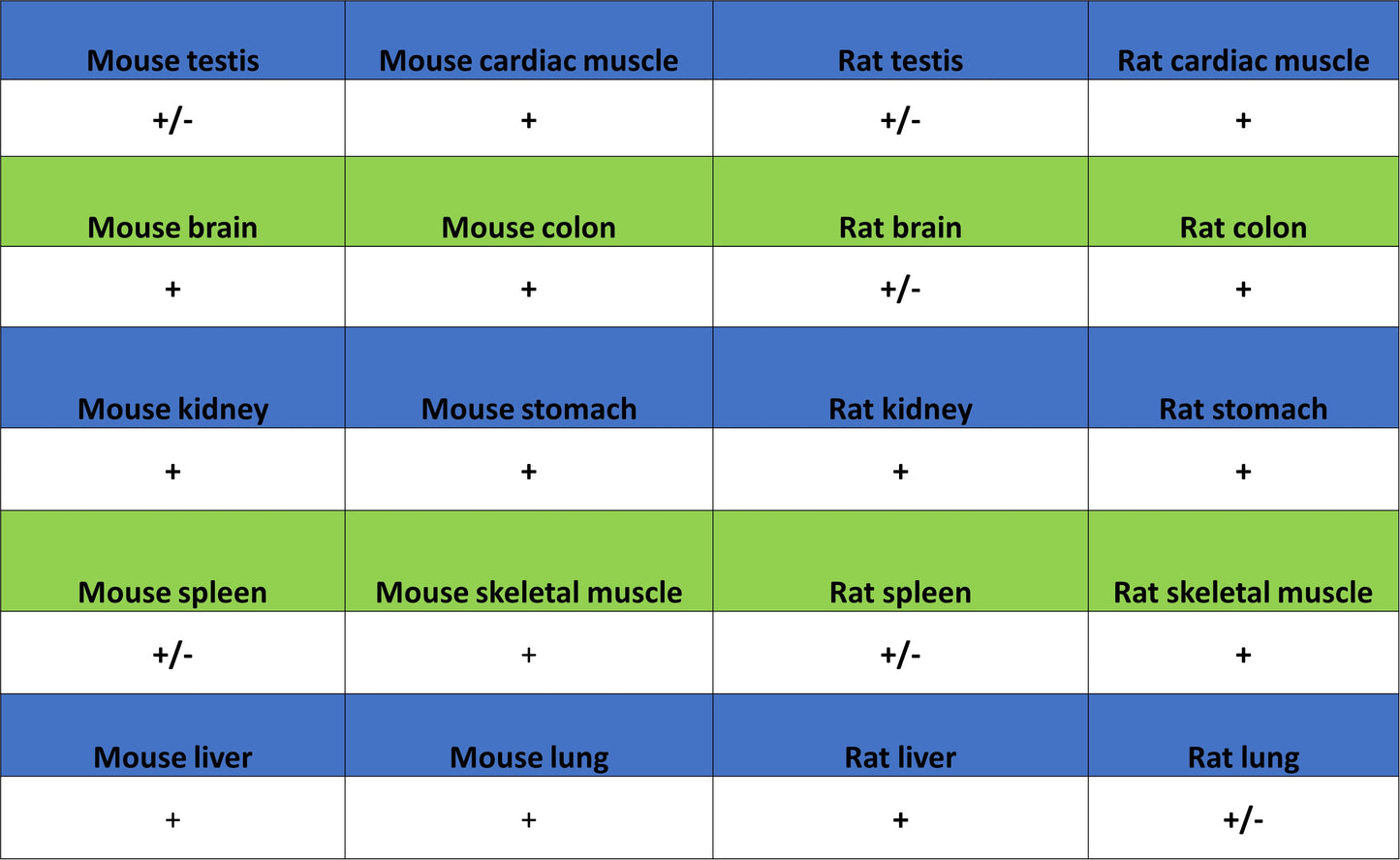
IHC shows positive staining in paraffin-embedded human cardiac muscle. Anti-NDUFB8 antibody was used at 1/100 dilution, followed by a HRP Polymer for Mouse IgG (ready to use). Counterstained with hematoxylin. Heat mediated antigen retrieval with Tris/EDTA buffer pH9.0 was performed before commencing with IHC staining protocol.
IHC shows positive staining in paraffin-embedded human skeletal muscle. Anti-NDUFB8 antibody was used at 1/100 dilution, followed by a HRP Polymer for Mouse IgG (ready to use). Counterstained with hematoxylin. Heat mediated antigen retrieval with Tris/EDTA buffer pH9.0 was performed before commencing with IHC staining protocol.