IHC shows positive staining in paraffin-embedded human kidney. Anti-Napsin A antibody was used at 1/1000 dilution, followed by a HRP Polymer for Mouse & Rabbit IgG (ready to use). Counterstained with hematoxylin. Heat mediated antigen retrieval with Tris/EDTA buffer pH9.0 was performed before commencing with IHC staining protocol.
Napsin A Recombinant Mouse mAb,PBS Only (SDT-4204)
Napsin A Recombinant Mouse mAb,PBS Only (SDT-4204)
Price:
Regular price
$200 USD
Regular price
Sale price
$200 USD
Unit price
per
For shipping services or bulk orders, you may request a quotation.
Secure checkout with

View full details
Product Details
Product Details
Product Specification
| Host | Mouse |
| Antigen | Napsin A |
| Synonyms | Napsin-A; Aspartyl protease 4 (ASP4; Asp 4); Napsin-1; TA01/TA02; NAP1; NAPA; NAPSA |
| Location | Secreted |
| Accession | O96009 |
| Clone Number | SDT-4204 |
| Antibody Type | Mouse mAb |
| Isotype | IgG1,k |
| Application | WB, IHC-P |
| Reactivity | Hu, Ms, Rt |
| Purification | Protein G |
| Concentration | 1 mg/ml |
| Conjugation | Unconjugated |
| Physical Appearance | Liquid |
| Storage Buffer | PBS |
| Stability & Storage | 12 months from date of receipt / reconstitution, -4 °C as supplied |
Dilution
| application | dilution | species |
| IHC-P | 1:1000 | Hu, Ms, Rt |
Background
Napsin A is a 38-kDa, pepsin-fold aspartic protease encoded by chromosome 19q13 and expressed almost exclusively in type II pneumocytes and renal proximal tubular epithelial cells, where it participates in surfactant protein B maturation and endolysosomal protein catabolism; immunohistochemically its strong, granular cytoplasmic staining serves as a highly sensitive and specific marker for primary lung adenocarcinoma (positive in ~85 % of cases) while being consistently negative in squamous cell carcinoma, small-cell carcinoma, and most non-pulmonary adenocarcinomas except renal clear-cell carcinoma, making it a key component of modern diagnostic panels alongside TTF-1 for distinguishing pulmonary origin in metastatic settings.
Picture
Picture
Immunohistochemistry
IHC shows positive staining in paraffin-embedded human lung. Anti-Napsin A antibody was used at 1/1000 dilution, followed by a HRP Polymer for Mouse & Rabbit IgG (ready to use). Counterstained with hematoxylin. Heat mediated antigen retrieval with Tris/EDTA buffer pH9.0 was performed before commencing with IHC staining protocol.
IHC shows positive staining in paraffin-embedded human lung adenocarcinoma. Anti-Napsin A antibody was used at 1/1000 dilution, followed by a HRP Polymer for Mouse & Rabbit IgG (ready to use). Counterstained with hematoxylin. Heat mediated antigen retrieval with Tris/EDTA buffer pH9.0 was performed before commencing with IHC staining protocol.
Negative control: IHC shows negative staining in paraffin-embedded human lung squamous cell carcinoma. Anti-Napsin A antibody was used at 1/1000 dilution, followed by a HRP Polymer for Mouse & Rabbit IgG (ready to use). Counterstained with hematoxylin. Heat mediated antigen retrieval with Tris/EDTA buffer pH9.0 was performed before commencing with IHC staining protocol.
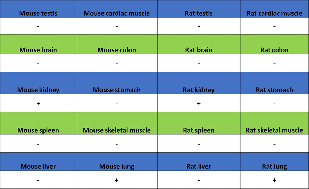
IHC shows positive staining in paraffin-embedded mouse kidney. Anti-Napsin A antibody was used at 1/1000 dilution, followed by a HRP Polymer for Mouse & Rabbit IgG (ready to use). Counterstained with hematoxylin. Heat mediated antigen retrieval with Tris/EDTA buffer pH9.0 was performed before commencing with IHC staining protocol.
IHC shows positive staining in paraffin-embedded mouse lung. Anti-Napsin A antibody was used at 1/1000 dilution, followed by a HRP Polymer for Mouse & Rabbit IgG (ready to use). Counterstained with hematoxylin. Heat mediated antigen retrieval with Tris/EDTA buffer pH9.0 was performed before commencing with IHC staining protocol.
IHC shows positive staining in paraffin-embedded rat kidney. Anti-Napsin A antibody was used at 1/1000 dilution, followed by a HRP Polymer for Mouse & Rabbit IgG (ready to use). Counterstained with hematoxylin. Heat mediated antigen retrieval with Tris/EDTA buffer pH9.0 was performed before commencing with IHC staining protocol.
IHC shows positive staining in paraffin-embedded rat lung. Anti-Napsin A antibody was used at 1/1000 dilution, followed by a HRP Polymer for Mouse & Rabbit IgG (ready to use). Counterstained with hematoxylin. Heat mediated antigen retrieval with Tris/EDTA buffer pH9.0 was performed before commencing with IHC staining protocol.