WB result of MYST2 Recombinant Rabbit mAb
Primary antibody: MYST2 Recombinant Rabbit mAb at 1/1000 dilution
Lane 1: SK-BR-3 whole cell lysate 20 μg
Lane 2: Caco-2 whole cell lysate 20 μg
Lane 3: MCF7 whole cell lysate 20 μg
Secondary antibody: Goat Anti-Rabbit IgG (H+L), HRP conjugated at 1/10000 dilution
Predicted MW: 70 kDa
Observed MW: 80 kDa
MYST2 Recombinant Rabbit mAb (S-3730-123)
MYST2 Recombinant Rabbit mAb (S-3730-123)
Price:
Regular price
$100 USD
Regular price
Sale price
$100 USD
Unit price
per
For shipping services or bulk orders, you may request a quotation.
Secure checkout with

View full details
Product Details
Product Details
Product Specification
| Host | Rabbit |
| Antigen | MYST2 |
| Synonyms | Histone acetyltransferase KAT7; Histone acetyltransferase binding to ORC1; Lysine acetyltransferase 7; MOZ; YBF2/SAS3; SAS2 and TIP60 protein 2 (MYST-2); HBO1 s; HBOa; KAT7 |
| Immunogen | Recombinant Protein |
| Location | Cytoplasm, Nucleus |
| Accession | O95251 |
| Clone Number | S-3730-123 |
| Antibody Type | Recombinant mAb |
| Isotype | IgG |
| Application | WB, ICC |
| Reactivity | Hu, Ms |
| Positive Sample | SK-BR-3, Caco-2, MCF7, F9, mouse testis |
| Purification | Protein A |
| Concentration | 0.5 mg/ml |
| Conjugation | Unconjugated |
| Physical Appearance | Liquid |
| Storage Buffer | PBS, 40% Glycerol, 0.05% BSA, 0.02% sodium azide |
| Stability & Storage | 12 months from date of receipt / reconstitution, -20 °C as supplied |
Dilution
| application | dilution | species |
| WB | 1:1000-1:5000 | Hu, Ms |
| ICC | 1:100-1:500 | Hu, Ms |
Background
MYST2 (MYST histone acetyltransferase 2), also known as KAT7 (Lysine Acetyltransferase 7) or HBO1 (Histone acetyltransferase binding to ORC1), is an evolutionarily highly conserved histone acetyltransferase belonging to the MYST family. It catalyzes the acetylation of lysine residues on histones H3 and H4 (particularly H3K14 and H4K8) via its MYST domain, thereby regulating chromatin accessibility and gene transcriptional activity. During the cell cycle, MYST2 serves as a core component of the pre-replicative complex, interacting with proteins such as ORC (Origin Recognition Complex) and promoting the licensing of DNA replication origins and the smooth progression of replication forks through histone acetylation, making it a critical enzyme for ensuring normal cell cycle progression and genomic stability. Its functional dysregulation is closely associated with tumor development and progression: MYST2 is aberrantly overexpressed in various cancers (such as breast cancer, liver cancer, and colorectal cancer), where it promotes tumor growth by activating pro-proliferative genes and anti-apoptotic signaling pathways, and its expression level is positively correlated with poor patient prognosis, positioning it as a promising anticancer therapeutic target. Furthermore, MYST2 is also involved in the self-renewal and differentiation regulation of embryonic stem cells, and its aberrant activity has also been found to be associated with certain developmental defects and neurological disorders, demonstrating its multifaceted importance in developmental biology and disease therapy.
Picture
Picture
Western Blot
WB result of MYST2 Recombinant Rabbit mAb
Primary antibody: MYST2 Recombinant Rabbit mAb at 1/1000 dilution
Lane 1: F9 whole cell lysate 20 μg
Lane 2: mouse teatis lysate 20 μg
Secondary antibody: Goat Anti-Rabbit IgG (H+L), HRP conjugated at 1/10000 dilution
Predicted MW: 70 kDa
Observed MW: 80 kDa
Immunocytochemistry
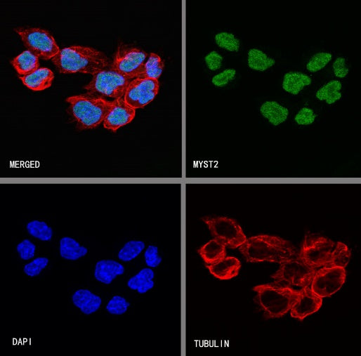
ICC shows positive staining in MCF7 cells. Anti-MYST2 antibody was used at 1/500 dilution (Green) and incubated overnight at 4°C. Goat polyclonal Antibody to Rabbit IgG - H&L (Alexa Fluor® 488) was used as secondary antibody at 1/1000 dilution. The cells were fixed with 100% ice-cold methanol and permeabilized with 0.1% PBS-Triton X-100. Nuclei were counterstained with DAPI (Blue). Counterstain with tubulin (Red).
ICC shows positive staining in F9 cells. Anti-MYST2 antibody was used at 1/100 dilution (Green) and incubated overnight at 4°C. Goat polyclonal Antibody to Rabbit IgG - H&L (Alexa Fluor® 488) was used as secondary antibody at 1/1000 dilution. The cells were fixed with 4% PFA and permeabilized with 0.1% PBS-Triton X-100. Nuclei were counterstained with DAPI (Blue). Counterstain with tubulin (Red).