WB result of MYH7 Recombinant Rabbit mAb
Primary antibody: MYH7 Recombinant Rabbit mAb at 1/1000 dilution
Lane 1: mouse heart lysate 20 µg
Lane 2: mouse skeletal muscle lysate 20 µg
Secondary antibody: Goat Anti-Rabbit IgG (H+L), HRP conjugated at 1/10000 dilution
Predicted MW: 223 kDa
Observed MW: 170-260 kDa
MYH7 Recombinant Rabbit mAb (S-4468-32)
MYH7 Recombinant Rabbit mAb (S-4468-32)
Price:
Regular price
$100 USD
Regular price
Sale price
$100 USD
Unit price
per
For shipping services or bulk orders, you may request a quotation.
Secure checkout with

View full details
Product Details
Product Details
Product Specification
| Host | Rabbit |
| Antigen | MYH7 |
| Synonyms | Myosin-7; Myosin heavy chain 7; Myosin heavy chain slow isoform (MyHC-slow); Myosin heavy chain; cardiac muscle beta isoform (MyHC-beta); MYHCB |
| Immunogen | Recombinant Protein |
| Location | Cytoplasm |
| Accession | P12883 |
| Clone Number | S-4468-32 |
| Antibody Type | Recombinant mAb |
| Isotype | IgG |
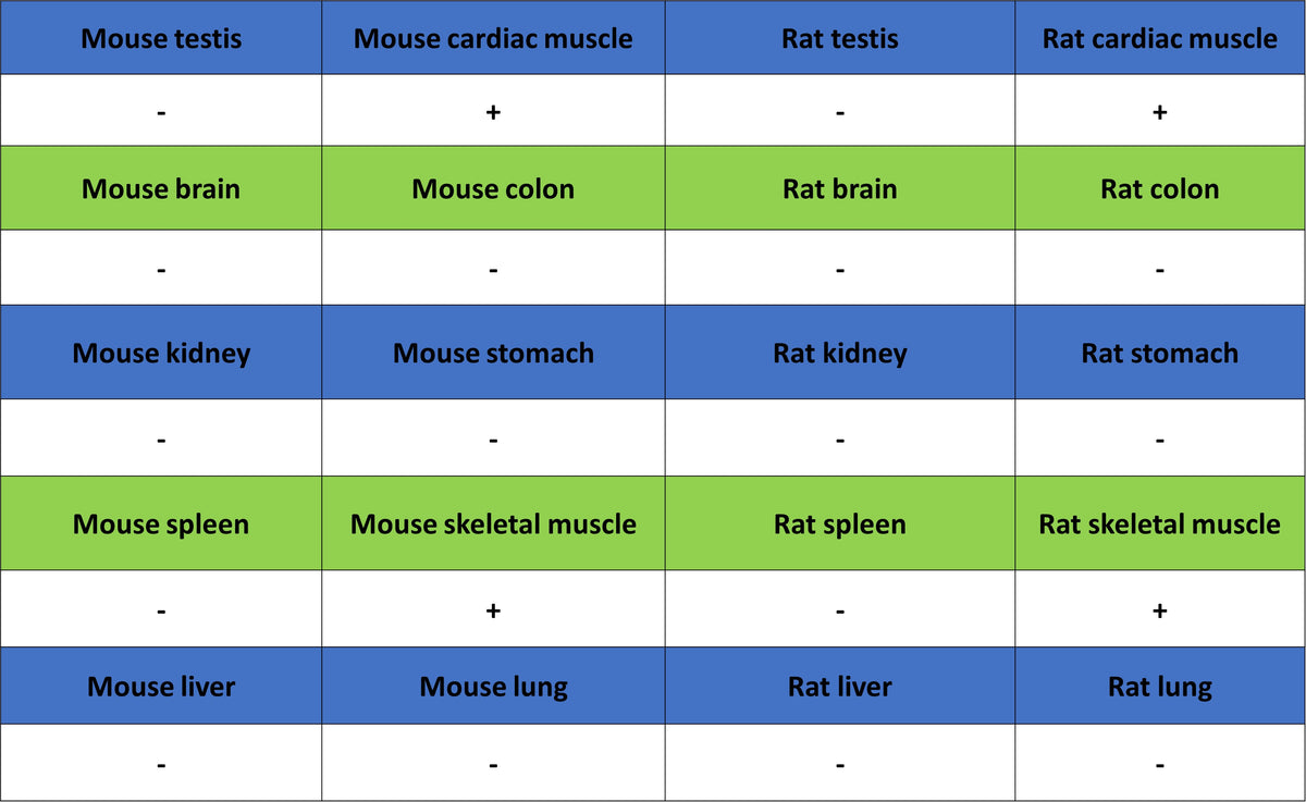
| Application | WB, IHC-P, IF |
| Reactivity | Hu, Ms, Rt |
| Positive Sample | mouse heart muscle, mouse skeletal muscle, rat heart muscle, rat skeletal muscle |
| Purification | Protein A |
| Concentration | 0.5 mg/ml |
| Conjugation | Unconjugated |
| Physical Appearance | Liquid |
| Storage Buffer | PBS, 40% Glycerol, 0.05% BSA, 0.02% sodium azide |
| Stability & Storage | 12 months from date of receipt / reconstitution, -20 °C as supplied |
Dilution
| application | dilution | species |
| WB | 1:1000-1:10000 | Ms, Rt |
| IHC-P | 1:2000 | Hu, Ms |
| ICC | 1:500 | Hu |
Background
MYH7, the slow/cardiac β-myosin heavy chain encoded by the MYH7 gene on chromosome 14q12, is the dominant motor protein of the human ventricle and type I skeletal muscle fibers, forming the hexameric thick-filament complex (two MYH7, two ELC, two RLC) whose ATP-driven conformational cycles generate ≈2 pN of force at 5–10 nm steps; pathogenic missense or truncating variants disrupt this chemomechanical cycle and the accompanying α-helical coiled-coil packing, producing a dosage-sensitive spectrum of disease—hypertrophic and dilated cardiomyopathy, myosin storage myopathy, Laing distal myopathy—characterized by hypertrophic sarcomeres, replacement fibrosis, and progressive heart or skeletal muscle failure, while physiologically MYH7 expression is induced by thyroid hormone, exercise, and β-adrenergic signaling that up-regulate slow oxidative programs, making the protein a central therapeutic target for small-molecule myosin inhibitors (mavacamten) and gene-editing strategies aimed at normalizing contractile kinetics and Ca²⁺ sensitivity.
Picture
Picture
Western Blot
WB result of MYH7 Recombinant Rabbit mAb
Primary antibody: MYH7 Recombinant Rabbit mAb at 1/1000 dilution
Lane 1: rat heart lysate 20 µg
Lane 2: rat skeletal muscle lysate 20 µg
Secondary antibody: Goat Anti-Rabbit IgG (H+L), HRP conjugated at 1/10000 dilution
Predicted MW: 223 kDa
Observed MW: 170-260 kDa
Immunohistochemistry
IHC shows positive staining in paraffin-embedded human cardiac muscle. Anti-MYH7 antibody was used at 1/2000 dilution, followed by a HRP Polymer for Rabbit IgG (ready to use). Counterstained with hematoxylin. Heat mediated antigen retrieval with Tris/EDTA buffer pH9.0 was performed before commencing with IHC staining protocol.
IHC shows positive staining in paraffin-embedded human skeletal muscle. Anti-MYH7 antibody was used at 1/2000 dilution, followed by a HRP Polymer for Rabbit IgG (ready to use). Counterstained with hematoxylin. Heat mediated antigen retrieval with Tris/EDTA buffer pH9.0 was performed before commencing with IHC staining protocol.
IHC shows positive staining in paraffin-embedded mouse skeletal muscle. Anti-MYH7 antibody was used at 1/2000 dilution, followed by a HRP Polymer for Rabbit IgG (ready to use). Counterstained with hematoxylin. Heat mediated antigen retrieval with Tris/EDTA buffer pH9.0 was performed before commencing with IHC staining protocol.
IHC shows positive staining in paraffin-embedded rat skeletal muscle. Anti-MYH7 antibody was used at 1/2000 dilution, followed by a HRP Polymer for Rabbit IgG (ready to use). Counterstained with hematoxylin. Heat mediated antigen retrieval with Tris/EDTA buffer pH9.0 was performed before commencing with IHC staining protocol.
Immunofluorescence
IF shows positive staining in paraffin-embedded human cardiac muscle. Anti-MYH7 antibody was used at 1/500 dilution (Green) and incubated overnight at 4°C. Goat polyclonal Antibody to Rabbit IgG - H&L (Alexa Fluor® 488) was used as secondary antibody at 1/1000 dilution. Counterstained with DAPI (Blue). Heat mediated antigen retrieval with EDTA buffer pH9.0 was performed before commencing with IF staining protocol.
IF shows positive staining in paraffin-embedded human skeletal muscle. Anti-MYH7 antibody was used at 1/500 dilution (Green) and incubated overnight at 4°C. Goat polyclonal Antibody to Rabbit IgG - H&L (Alexa Fluor® 488) was used as secondary antibody at 1/1000 dilution. Counterstained with DAPI (Blue). Heat mediated antigen retrieval with EDTA buffer pH9.0 was performed before commencing with IF staining protocol.