WB result of MYBPC3 Recombinant Rabbit mAb
Primary antibody: MYBPC3 Recombinant Rabbit mAb at 1/1000 dilution
Lane 1: mouse heart lysate 20 µg
Secondary antibody: Goat Anti- rabbit IgG, (H+L), HRP conjugated at 1/10000 dilution
Predicted MW: 140 kDa
Observed MW: 150 kDa
MYBPC3 Recombinant Rabbit mAb (S-5476)
MYBPC3 Recombinant Rabbit mAb (S-5476)
Price:
Regular price
$100 USD
Regular price
Sale price
$100 USD
Unit price
per
For shipping services or bulk orders, you may request a quotation.
Secure checkout with

View full details
Product Details
Product Details
Product Specification
| Host | Rabbit |
| Antigen | MYBPC3 |
| Synonyms | Myosin-binding protein C, cardiac-type |
| Accession | Q14896 |
| Clone Number | S-5476 |
| Antibody Type | Recombinant mAb |
| Isotype | IgG |
| Application | WB, IHC-P |
| Reactivity | Hu, Ms |
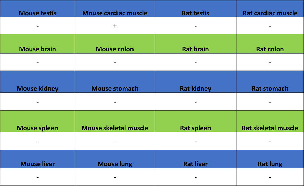
| Positive Sample | mouse heart, rat heart |
| Purification | Protein A |
| Concentration | 0.5 mg/ml |
| Conjugation | Unconjugated |
| Physical Appearance | Liquid |
| Storage Buffer | PBS, 40% Glycerol, 0.05% BSA, 0.02% sodium azide |
| Stability & Storage | 12 months from date of receipt / reconstitution, -20 °C as supplied |
Dilution
| application | dilution | species |
| WB | 1:1000 | Hu, Ms |
| IHC-P | 1:500 | Hu, Ms |
Background
MYBPC3 is a key thick filament‑associated protein located in the cardiac sarcomere. This protein is predominantly and specifically expressed in cardiac muscle and contains multiple immunoglobulin‑like and fibronectin type III domains. Through interactions with various sarcomeric components—including myosin, actin, and titin—it finely regulates the speed and force of myocardial contraction. Notably, MYBPC3 is one of the most important causative genes for familial hypertrophic cardiomyopathy (HCM), accounting for approximately 30%–40% of all HCM cases. Among these mutations, truncating variants (such as nonsense and frameshift mutations) are the most common, typically leading to reduced expression of cMyBP‑C protein (haploinsufficiency), which destabilizes sarcomeric structure and function and results in myocardial pathology characterized by asymmetric septal hypertrophy. Although most carriers of MYBPC3 mutations present with later onset and milder symptoms, specific mutation types can also cause early‑onset, severe hypertrophy and heart failure. The clinical phenotype is highly heterogeneous, ranging from completely asymptomatic to sudden cardiac death.
Picture
Picture
Western Blot
WB result of MYBPC3 Recombinant Rabbit mAb
Primary antibody: MYBPC3 Recombinant Rabbit mAb at 1/1000 dilution
Lane 1: rat heart lysate 20 µg
Secondary antibody: Goat Anti- rabbit IgG, (H+L), HRP conjugated at 1/10000 dilution
Predicted MW: 140 kDa
Observed MW: 150 kDa
Immunohistochemistry
IHC shows positive staining in paraffin-embedded human cardiac muscle. Anti-MYBPC3 antibody was used at 1/500 dilution, followed by a HRP Polymer for Rabbit IgG (ready to use). Counterstained with hematoxylin. Heat mediated antigen retrieval with Tris/EDTA buffer pH9.0 was performed before commencing with IHC staining protocol.
Negative control: IHC shows negative staining in paraffin-embedded human skeletal muscle. Anti-MYBPC3 antibody was used at 1/500 dilution, followed by a HRP Polymer for Rabbit IgG (ready to use). Counterstained with hematoxylin. Heat mediated antigen retrieval with Tris/EDTA buffer pH9.0 was performed before commencing with IHC staining protocol.
Negative control: IHC shows negative staining in paraffin-embedded human colon. Anti-MYBPC3 antibody was used at 1/500 dilution, followed by a HRP Polymer for Rabbit IgG (ready to use). Counterstained with hematoxylin. Heat mediated antigen retrieval with Tris/EDTA buffer pH9.0 was performed before commencing with IHC staining protocol.
IHC shows positive staining in paraffin-embedded mouse cardiac muscle. Anti-MYBPC3 antibody was used at 1/500 dilution, followed by a HRP Polymer for Rabbit IgG (ready to use). Counterstained with hematoxylin. Heat mediated antigen retrieval with Tris/EDTA buffer pH9.0 was performed before commencing with IHC staining protocol.
Negative control: IHC shows negative staining in paraffin-embedded mouse skeletal muscle. Anti-MYBPC3 antibody was used at 1/500 dilution, followed by a HRP Polymer for Rabbit IgG (ready to use). Counterstained with hematoxylin. Heat mediated antigen retrieval with Tris/EDTA buffer pH9.0 was performed before commencing with IHC staining protocol.