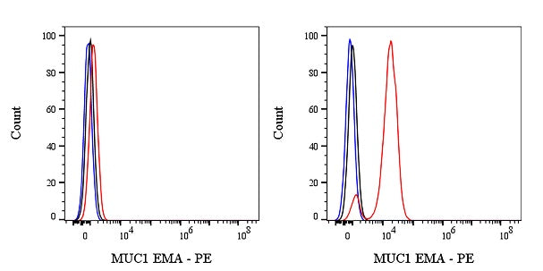
Flow cytometric analysis of HCT116 (Human colorectal carcinoma epithelial cell, left) / T-47D (Human ductal breast epithelial tumor epithelial cell, right) stained with PE Mouse IgG1, k Isotype Control (black line histogram) and SDT PE MUC1/EMA Mouse mAb (red line histogram) at 1/1000 dilution (0.1μg), cells without incubation with primary antibody and secondary antibody (blue line histogram) was used as unlabeled control. Flow cytometry and data analysis were performed using Agilent NovoCyte Quanteon and FlowJo™ software.
Negative control: HCT116
MUC1/EMA Mouse mAb (PE Conjugate) (S-777-37)
MUC1/EMA Mouse mAb (PE Conjugate) (S-777-37)
Price:
Regular price
$185 USD
Regular price
Sale price
$185 USD
Unit price
per
For shipping services or bulk orders, you may request a quotation.
Secure checkout with

View full details
Product Details
Product Details
Product Specification
| Host | Mouse |
| Synonyms | Mucin-1; Breast carcinoma-associated antigen DF3; Cancer antigen 15-3 (CA 15-3); Carcinoma-associated mucin; Episialin; H23AG; CD227; PUM |
| Immunogen | Synthetic Peptide |
| Location | Cytoplasm, Cell membrane |
| Accession | P15941 |
| Clone Number | S-777-37 |
| Antibody Type | Mouse mAb |
| Isotype | IgG1,k |
| Application | ICC, IF, ICFCM |
| Reactivity | Hu, Rt |
| Purification | Protein G |
| Concentration | 1 mg/ml |
| Conjugation | PE |
| Physical Appearance | Liquid |
| Storage Buffer | PBS, 1% BSA, 0.3% Proclin 300 |
| Stability & Storage | 12 months from date of receipt / reconstitution, 2 to 8 °C as supplied |
Dilution
| application | dilution | species |
| ICC | 1:500 | Hu |
| IF | 1:200 | Hu, Rt |
| ICFCM | 1:1000 | Hu |
Background
MUC1, also historically known as Epithelial Membrane Antigen (EMA), is a large transmembrane glycoprotein belonging to the mucin family that is normally expressed on the apical surface of most secretory epithelial cells, where its heavily O-glycosylated extracellular domain serves critical protective and lubricating functions while maintaining cell polarity; however, in malignant transformations across various carcinomas (including breast, lung, pancreatic, and ovarian cancers), MUC1 undergoes profound aberrations characterized by underglycosylation, loss of polarized distribution, and overexpression, which expose novel peptide epitopes and facilitate tumor progression through mechanisms such as shielding cancer cells from immune surveillance, promoting metastasis via interactions with selectins, and activating oncogenic signaling pathways like NF-κB and β-catenin, thereby making it not only a widely used diagnostic immunohistochemical marker for epithelial-derived tumors but also a prominent target for therapeutic strategies including monoclonal antibodies, antibody-drug conjugates, and cancer vaccines.
Picture
Picture
FC
Immunocytochemistry
ICC shows positive staining in T-47D cells (top panel) and negative staining in HCT116 cells (below panel). Anti-MUC1/EMA (PE Conjugate) antibody was used at 1/500 dilution (Red) and incubated overnight at 4°C. The cells were fixed with 4% PFA and permeabilized with 0.1% PBS-Triton X-100. Nuclei were counterstained with DAPI (Blue).
Immunofluorescence
IF shows positive staining in paraffin-embedded human colon. Anti-MUC1/EMA (PE Conjugate) antibody was used at 1/200 dilution (Red) and incubated overnight at 4°C. Counterstained with DAPI (Blue). Heat mediated antigen retrieval with EDTA buffer pH9.0 was performed before commencing with IF staining protocol.
IF shows positive staining in paraffin-embedded rat stomach. Anti-MUC1/EMA (PE Conjugate) antibody was used at 1/200 dilution (Red) and incubated overnight at 4°C. Counterstained with DAPI (Blue). Heat mediated antigen retrieval with EDTA buffer pH9.0 was performed before commencing with IF staining protocol.