WB result of MTFR2 Recombinant Rabbit mAb
Primary antibody: MTFR2 Recombinant Rabbit mAb at 1/1000 dilution
Lane 1: HCT 116 whole cell lysate 20 µg
Lane 2: SW480 whole cell lysate 20 µg
Lane 3: T-47D whole cell lysate 20 µg
Lane 4: MDA-MB-231 whole cell lysate 20 µg
Lane 5: K562 whole cell lysate 20 µg
Lane 6: Jurkat whole cell lysate 20 µg
Lane 7: LoVo whole cell lysate 20 µg
Secondary antibody: Goat Anti- rabbit IgG, (H+L), HRP conjugated at 1/10000 dilution
Predicted MW: 43 kDa
Observed MW: 40 kDa
MTFR2 Recombinant Rabbit mAb (S-3842-54)
MTFR2 Recombinant Rabbit mAb (S-3842-54)
Price:
Regular price
$100 USD
Regular price
Sale price
$100 USD
Unit price
per
For shipping services or bulk orders, you may request a quotation.
Secure checkout with

View full details
Product Details
Product Details
Product Specification
| Host | Rabbit |
| Antigen | MTFR2 |
| Synonyms | Mitochondrial fission regulator 2; DUFD1; FAM54A |
| Immunogen | Synthetic Peptide |
| Location | Mitochondrion |
| Accession | Q6P444 |
| Clone Number | S-3842-54 |
| Antibody Type | Recombinant mAb |
| Isotype | IgG |
| Application | WB |
| Reactivity | Hu, Ms, Rt |
| Positive Sample | HCT116, SW480, T-47D, MDA-MB-231, K562, Jurkat, LoVo, mouse testis, rat testis |
| Purification | Protein A |
| Concentration | 0.5 mg/ml |
| Conjugation | Unconjugated |
| Physical Appearance | Liquid |
| Storage Buffer | PBS, 40% Glycerol, 0.05% BSA, 0.03% Proclin 300 |
| Stability & Storage | 12 months from date of receipt / reconstitution, -20 °C as supplied |
Dilution
| application | dilution | species |
| WB | 1:1000-1:5000 | Hu, Ms, Rt |
Background
MTFR2 (Mitochondrial Fission Regulator 2), also known as MFF or sometimes referred to in specific contexts alongside related fission factors, is a critical outer mitochondrial membrane protein that acts as a primary receptor for recruiting the dynamin-related GTPase Drp1 (Dynamin-related protein 1) to facilitate mitochondrial fission, a fundamental process required for maintaining mitochondrial morphology, distribution, and quality control within eukaryotic cells. Located on the cytosolic face of the outer mitochondrial membrane, MTFR2 contains specific domains that directly interact with Drp1 oligomers, effectively anchoring the fission machinery at designated constriction sites where it promotes GTP hydrolysis-driven membrane scission, thereby enabling the division of elongated mitochondrial networks into smaller fragments necessary for cellular functions such as mitosis, apoptosis, and the isolation of damaged mitochondrial segments for subsequent mitophagy. Dysregulation of MTFR2 expression or function can lead to aberrant mitochondrial hyperfusion or excessive fragmentation, which has been implicated in various pathological conditions including neurodegenerative diseases, metabolic disorders, and cancer, highlighting its essential role as a key molecular switch that balances mitochondrial dynamics in response to cellular energy demands and stress signals.
Picture
Picture
Western Blot
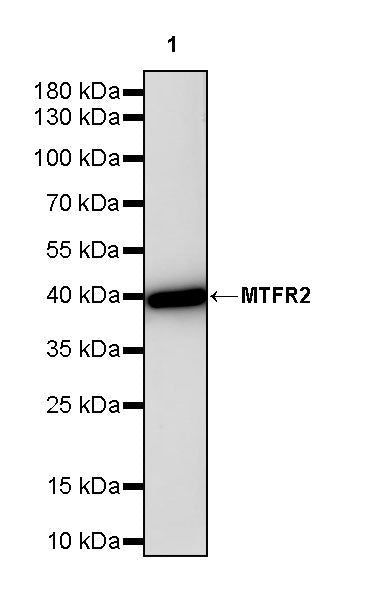
WB result of MTFR2 Recombinant Rabbit mAb
Primary antibody: MTFR2 Recombinant Rabbit mAb at 1/1000 dilution
Lane 1: mouse testis lysate 20 µg
Secondary antibody: Goat Anti- rabbit IgG, (H+L), HRP conjugated at 1/10000 dilution
Predicted MW: 43 kDa
Observed MW: 40 kDa
WB result of MTFR2 Recombinant Rabbit mAb
Primary antibody: MTFR2 Recombinant Rabbit mAb at 1/1000 dilution
Lane 1: rat testis lysate 20 µg
Secondary antibody: Goat Anti- rabbit IgG, (H+L), HRP conjugated at 1/10000 dilution
Predicted MW: 43 kDa
Observed MW: 40 kDa