
Mouse TL ELISA Kit
Mouse TL ELISA Kit
Price:
Regular price
$508 USD
Regular price
Sale price
$508 USD
Unit price
per
For shipping services or bulk orders, you may request a quotation.
Secure checkout with

View full details
Product Details
Product Details
Product Specification
| Usage |
1. Self-prepared test equipment required for the experiment 1 Microplate reader, ( wavelength 450nm Optical filter ) 2 、 37°C Incubator ( Cell use is not recommended CO2 Incubator ) 3 , automatic plate washer or multi-channel pipette /5ml Dropper ( For manual plate washing ) 4 , precise single channel (0.5-10μL,5-50μL, 20-200μL, 200-1000μL) And multi-channel pipettes ( Pipette needs to be calibrated before use ) 。 5 Sterile, EP Tubes and disposable tips 6 , absorbent paper and sample loading tank 7 Deionized water or distilled water 2. Sample processing and requirements: Serum: Whole Blood Samples Hold at Room Temperature 2 Hour or 2-8°C Overnight. 1000×g Centrifugation 20 Minutes, take the supernatant. Can be tested immediately, or frozen in packages according to one-time usage -20°C Or -80°C 。 Plasma: Anticoagulants recommended EDTA-Na2/K2 , after sample collection 30 Within minutes 2-8°C , 1000×g Centrifugation 15 Minutes, take the supernatant. Can be tested immediately, or frozen in packages according to one-time usage -20°C Or -80°C 。 Tissue samples: Tissue samples are generally made into tissue homogenates, and the processing methods are as follows: 1 Placing the target tissue on ice, using pre-cooled PBS Buffer (0.01M , pH=7.4) Wash to remove residual blood, weigh and use later. 2 Grinding the tissue homogenate with the lysate on ice. The volume of lysate added depends on the weight of the tissue. Generally, each 1g Tissue Fragment Use 9ml Lysate. It is also recommended to add protease inhibitors to the lysate, such as 1mMPMSF 。 3 Can be reused for ultrasonic crushing or repeated freezing and thawing for further treatment ( In the process of ultrasonic crushing, ice bath is needed to cool down; Repeated freeze-thaw method can be repeated 2 Time ) 。 4 Dissolving the prepared homogenate in 5000×g Centrifugation 5 Minutes, retain the supernatant for detection. Or frozen in packages according to one-time usage -20°C Or -80°C 。 5 According to the experimental needs, the total protein concentration of the tissue homogenate sample can be measured first , To facilitate data analysis, recommended BCA Law. The total protein concentration is generally adjusted to 13mg/ml Used for ELISA Detection. Certain tissue samples, such as liver, kidney, and pancreas, contain higher concentrations of endogenous peroxygenase , When the sample concentration is high, it will react with the chromogenic substrate, resulting in false positives. Can try to use 1%H2O2 Inactivation 15min Re-test. Note: Commonly used lysate PBS Buffer, or use moderate strength RIPA Lysate. Using RIPA When lysing solution, PH The value needs to be adjusted to PH7.3 , avoid using containing NP-40 , Triton X-100 And DTT The components will seriously inhibit the work of the kit. Recommended use 50mM Tris+0.9%NaCL+0.1% SDS,PH 7.3 , can be prepared by yourself or contact us to purchase. Cell culture supernatant: Collecting the supernatant, 2-8°C , 2500rpm Centrifugation 5min , the clarified cell culture supernatant was collected. Use immediately for testing, or dispense in a single-use amount -80°C Freeze for later use. Cell lysate: 1 Collection and lysis of suspended cells: 2-8°C , 2500rpm Centrifugation 5min , collecting the cells. Add the pre-cooled PBS Gently mix and clean, 2-8°C , 2500rpm Centrifugation 5min , collecting the cells. join 0.5-1ml Cell lysate and appropriate amount of protease inhibitor ( Such as PMSF , working concentration 1mmol/L) , placed on ice, lysed 30min-1h , Or cooperate with ultrasonic crushing. 2 , collection and lysis of adherent cells: aspirate the supernatant and add pre-cooled PBS Wash three times. join 0.5-1ml Cell lysate and appropriate amount of protease inhibitor ( Such as PMSF , working concentration 1mmol/L) , gently scrape off the adherent cells with a cell scraper. The cell suspension was transferred to a centrifuge tube, placed on ice, and lysed 30min-1h , or cooperate with ultrasonic crushing. 3 During the cell lysis process, the centrifuge tube can be blown with the gun tip or shaken intermittently to fully lyse the protein , Appears slimy yes DNA , can be disrupted using ultrasonic DNA 。 ( Or with ultrasound 3-5mm Probe, Power 150-300W, Sonicated samples on ice, working 1-2 Seconds, stop 30 Seconds, 3~5 A cycle. ) 4 Complete cracking or ultrasonic crushing, 2-8°C , 10000rpm Centrifugation 10min , the supernatant is moved into EP In tubes, immediately used for testing, or dispensed into one-time usage -80°C Freeze for later use. Note: The precautions are the same as for tissue samples. Other biological samples:2-8°C , 1000×g Centrifuge samples 20 Minutes. Collect the supernatant for immediate detection, or divide it into one-time use -80°C Freeze for later use. 3. Other precautions for samples 1 The test tube for collecting blood should be a disposable endotoxin-free test tube. Avoid the use of hemolyzed, hyperlipidemic samples. 2 Optimal storage conditions of samples: 2-8°C Storage should be less than 5 Day, -20°C Should not exceed 6 Months, -80°C Should not exceed 2 Years, it should be stored in liquid nitrogen beyond the above time. In order to reduce ice crystals when frozen specimens are thawed (0°C) Damage to the sample shall be adopted 15-25°C Quickly melt in water bath, centrifuge to remove the precipitate after melting, mix well and use for detection. 3 The detection range of the kit is not equivalent to the concentration range of the substance to be tested in the sample. If the concentration of the test substance in the sample is too high or too low, please dilute or concentrate the sample appropriately. 4 If the sample tested is special and there is no reference data, it is recommended to do pre-experiments to verify its effectiveness. 4. Sample dilution plan: If other dilution ratios are required for your model set samples, please refer to the general dilution protocol below ( This protocol is a dilution protocol that does not set up double wells for testing. When you need to set up a double hole, please add the sample and diluent volume x Number of complex holes ) : Dilution 2 Times (1/2) : One-step dilution. Take 60ul Sample addition 60ul In the sample dilution, mix gently. Dilution 5 Times (1/5) : One-step dilution. Take 24ul Sample addition 96ul In the sample dilution, mix gently. Dilution 10 Times (1/10) : One-step dilution. Take 12ul Sample addition 108ul In the sample dilution, mix gently. Dilution 20 Times (1/20) : One-step dilution. Take 6ul Sample addition 114ul In the sample dilution, mix gently. Dilution 50 Times (1/50) : One-step dilution. Take 3ul Samples and 47ul Normal saline ( I.e 0.9% Sodium chloride ) join 100ul In the sample dilution, mix gently. Dilution 100 Times (1/100) : One-step dilution. Take 3ul Samples and 177ul Normal saline addition 120ul In the sample dilution, mix gently. Dilution 1000 Times (1/1000) : Two-step dilution, you can dilute first 50 Times ( This step is entirely diluted with saline ) , re-dilution 20 Times. Gently mix well. Dilution 10000 Times (1/10000) : Two-step dilution, you can dilute first 100 Times ( This step is entirely diluted with saline ) , re-dilution 100 Times. Gently mix well. Dilution 100000 Times (1/100000) : Three-step dilution, you can dilute first 50 Times, re-dilute 20 Times ( The first two steps are all diluted with normal saline ) , final dilution 100 Times. Gently mix well. Note: The amount of liquid taken during each dilution step is not less than 3ul , the dilution factor is not more than 100 Times. Each step of dilution should be mixed evenly to avoid foaming. 5. Preparations before testing: ahead of schedule 20 Minutes remove the kit from the refrigerator and equilibrate to room temperature (18-25°C) 。 If the kit needs to be used multiple times, please take out only the enzyme-labeled slats and standards required for this experiment, and store the remaining enzyme-labeled slats and standards according to the specified conditions. 1 Lotion, : With deionized water or distilled water ( The recommended resistivity is 18MΩ Ultrapure water ) Will 30ml Concentrated wash (48T For 15ml) Diluted to 750ml(48T For 375ml) And mix well. Or according to the requirements of the experiment, take an appropriate amount of concentrated washing liquid and dilute to 25 Double volume and mix well, return the unused solution 2-8°C 。 If crystals are formed in the concentrated washing liquid, they may be 40°C Heating in water bath ( The heating temperature should not exceed 50°C) , until the crystals are completely dissolved, mix well and use. The prepared lotion is best used on the same day. If it is not used up, it can be stored in 2-8°C , not more than 48 Hours. 2 Standard, : 2.1 , control the freeze-dried standard quality in 10000×g Centrifugation 1 Minutes. Marked as Zerotube. 2.2 Take 1ml Add the sample diluent to the lyophilized standard tube, screw the tube cap tightly, and let it stand at room temperature 2 Minutes, upside down several times and gently mix well ( Or join 1mL Sample dilution, stand 1-2 After minutes, mix well with a low speed vortex instrument 3-5 second ) 。 1000×g Low speed centrifugation 1 Minutes, collect the liquid to the bottom of the tube. 2.3 Gradient dilution: separately 7 1 EP Tubes, respectively labeled as 1/2 、 1/4 、 1/8 、 1/16 、 1/32 、 1/64 And blank 。 First in each EP Add separately into the tube 0.3ml Sample dilution of. Take again 0.3mlZero tube Standard solution is added to 1/2 Tube, mix thoroughly. Take again 0.3ml1/2 Tube standard solution to 1/4 Tube, mix thoroughly. Take again 0.3ml1/4 Tube standard solution to 1/8 Tube, mix thoroughly, and so on. attention BlankEP Only the sample dilution is in the tube. At this point, from Zerotube Tube to blank Take care of this 8 1 EP The concentration of the standard in the tube is 1000ng/ml,500ng/ml,250ng/ml, 125ng/ml,62.5ng/ml, 31.25ng/ml, 15.625ng/ml, 0ng/ml 。  Note : Dissolved tube zero standard, please store in 2-8°C , and in 12 Use within hours. For other diluted gradient standard working solution, please refer to 2 Use within hours. Before working fluid experiment 30 Ready within minutes, ready-to-use, not suitable for long-term storage. 3.1 , reconstitution: 2000×g Centrifugation 1 Minutes, the biotin will be concentrated - The antibody was collected to the bottom of the tube. Take purified water 70ul , added to the biotin-labeled antibody tube, completely dissolved After dissolving and mixing, 1000×g Low speed centrifugation 1 Minutes, placed in 2-8°C Save. 3.2 Calculate the total volume of working fluid required: 50ul/ Hole × Number of holes. ( It is best to prepare more than the total volume 100ul-200ul Amount of ) 3.3 , press with antibody dilution 1/100 Dilute concentrated biotin in proportion of - Antibody, mix well. ( If will 10ul Concentrated biotin - Antibody addition 990ul In antibody dilution ) 4 、 HRP- Streptavidin (SABC) Working fluid: Pre-experiment 30 Ready within minutes, ready-to-use, not suitable for long-term storage. 4.1 Calculate the total volume of working fluid required: 100ul/ Hole × Number of holes. ( It is best to prepare more than the total volume 100ul-200ul The amount of. ) 4.2 、 1000×g Low speed centrifugation 1 Minutes, will concentrate SABC Collect to the bottom of the tube. 4.3 Using SABC Diluent press 1/100 Dilution and concentration SABC , mix well. ( If will 10ul Concentration of SABC join 990ulSABC In diluent) 6. Operation steps: When diluting samples and reagents, they should be thoroughly mixed. It is recommended that a standard curve be plotted for each test. 1 Set the standard hole and the sample hole to be measured, and record their positions. In order to reduce experimental errors, it is recommended to set up double holes for standards and samples. Please wash the plate with wash buffer before loading 2 Times! 2 Sample addition: add to the corresponding well 50ul Standard or sample to be tested, then immediately add to each well 50ul Prepared biotin-labeled detection antibody working solution, gently shake the enzyme labeled plate and mix well 1 Minutes, apply the laminating film, and in 37°C Static incubation 45 Minutes. (Add the solution to the bottom of the microplate and avoid contact with the tube wall and blistering as much as possible.) 3 Washing plate 3 Second: Remove the coating film, suck or shake off the liquid in the enzyme labeled plate, and pat it on clean absorbent paper 2-3 Times. Add wash buffer per well 350ul , immersion 1 Minutes, discard the liquid in the hole and pat it on absorbent paper 2-3 Times. Repeat this plate washing step 3 Times. 4 Add HRP- Streptavidin (SABC) : Add to each well 100ulSABC Working fluid. Apply a laminating film, 37°C Static incubation 30 Minutes. ( At the same time the whole瓶 TMB Put in 37°C Equilibrium in incubator ) 5 Washing plate 5 Second: Remove the coating film and wash the plate with washing buffer 5 Time, method reference steps 3 。 6 Add TMB Chromogenic substrate: added to each well 90ulTMB Chromogenic substrate, coated with film, in 37°C Static incubation in the dark 10-20 Minutes. Turn on the plate reader to preheat 15min 。 ( Note: Do not use the formulation HRP Loading tank for the conjugate. Color development According to the actual change of color, the reaction time can be shortened or extended, but it cannot exceed 30 Minutes. When a better blue gradient appears in the standard well, the reaction can be terminated. The color rendering intensity is not easily too weak or too strong. 7 Add reaction stop solution: after color development, the liquid in the hole cannot be discarded, and add to each hole 50ul Reaction stop solution. The color will instantly change from blue to yellow. Sequence and addition of stop solution TMB The order of the substrates is the same. 8 、 OD Measurement of values: immediately use a microplate reader in 450nm Read at OD450 Numerical value. (If your microplate reader has a selectable correction wavelength, set to 570nm Or 630nm 。 The calibration reading is OD450 Minus the value of OD570 Or OD630 The value of. This method corrects and removes non-chromogenic substances OD Value, so as to obtain more accurate detection results. If the plate reader does not 570nm Or 630nm Wavelength, you can use the original OD450 Value.) VII. Calculation of experimental results: Results Calculation: 1 Take the average of standard and sample replica wells OD450 Value (using raw OD450 Value or correction read value). 2 , taking concentration as the abscissa, averaging OD450 Values are ordinates, and four-parameter equations can be used 4PL Draw the standard curve. You can also use the mapping software that comes with the plate reader ( Such as Thermo FC Model plate reader SkanIt RE Software ) Or Curve Expert 1.3 or 1.4 Professional Software ( Our website can be downloaded and used for free ) Draw the standard curve. 3 The sample OD450 By substituting the value into the standard curve, the concentration value of the sample can be calculated. If the sample has been diluted, multiply it by the corresponding dilution factor. Description of different methods for drawing standard curves: 1 , linear plot: one axis represents the concentration of antigen, the other represents the read OD450 Value. R2 Values are typically used here to determine the fit, values greater than 0.99 Indicates that the fit is very good. However, linear plots tend to compress the data points on the lower end of the curve, resulting in inaccurate calculation results. 2 , semi-logarithmic graph: helps to counteract the lower end compression caused by linear graph. Semi-logarithmic plots use the logarithm of concentration versus readings. This method usually results in a more uniform distribution of data points s Shape curve. 3 , logarithm / Double logarithmic plot: Provides good linearity for low to medium concentration range. But the high end of the range tends to lose linearity. 4 , four or five parameter equations (4PL Or 5PL) Curve: The method is more complex and takes into account other parameters, such as maximum and minimum values, so it requires more complex calculations. 4PL Assume symmetry around the inflection point, while 5PL Asymmetric cases are considered and are generally more suitable for immunoassays. If your software allows, then 4-PL And 5-PL Will apply to most ELISA Calibrate the standard curve. Experimental data and standard curve: This product has been tested by the quality control department and meets the performance requirements of the instruction manual. ( Laboratory humidity is 20%-60% , the temperature is 18°C-25°C 。 Before color development TMB Balance to 37°C After adding enzyme labeled plate wells, 37°C Incubate in the dark 15 Minutes. ) Due to differences in specific experimental environment and operation, the following experimental data and standard curves are for reference only, and experimenters need to establish standard curves according to their own experiments. 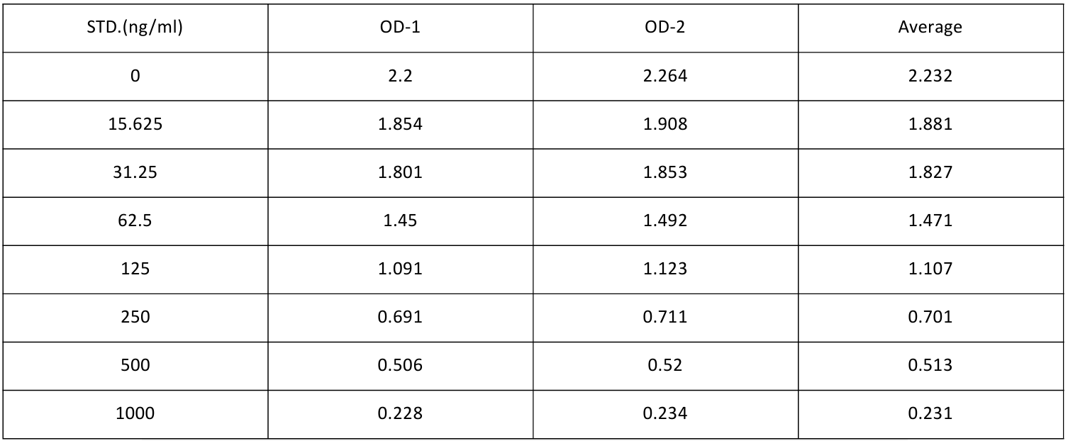  8. Kit performance: 1 Precision: In-plate precision: low, medium and high concentration samples are in the same 1 On-block plate detection 20 Times. Inter-plate precision: Low, medium and high concentration samples are respectively in 3 On-block plate detection 20 Times.
2 Recovery rate: Combine a certain amount of TC Added to the sample, and the measured values were added to the sample by comparing the measured values with TC The expected amounts of were compared to calculate the recovery.
3 Linear dilution: The appropriate concentration will be added TC The samples were diluted separately 2 Times, 4 Times, 8 Times to obtain the recovery range.
The unopened kit is available in 37°C And 2-8°C Stability experiments were performed and stability data were obtained.
|
||||||||||||||||||||||||||||||||||||||||||||||||||||||||||||||||||||||||||||
| Sensitivity | 9.375ng/ml | ||||||||||||||||||||||||||||||||||||||||||||||||||||||||||||||||||||||||||||
| Species Reactivity | Mouse | ||||||||||||||||||||||||||||||||||||||||||||||||||||||||||||||||||||||||||||
| Theory | This kit uses competitive ELISA assay, and the experimental duration is 2 hours. The plates supplied in the kit have been pre-coated with TC. The TC to be tested in the sample or standard will compete with the TC previously coated on the microplate for binding to a fixed amount of biotin-labeled anti-TC antibody. Free components are washed away. HRP-streptavidin (SABC) is added, and biotin specifically binds to streptavidin to form an immune complex. The unbound conjugate was washed off and TMB chromogenic substrate was added. The TMB became blue under the catalysis of horseradish peroxidase (HRP) and turned yellow after the reaction stop solution was added. The OD value was measured at 450nm with a microplate reader. Using curve equation software, the OD450 value of the sample was compared to the standard curve to determine the concentration of TC in the sample. There is an inverse proportion between the concentration of the target substance and the OD450 value. | ||||||||||||||||||||||||||||||||||||||||||||||||||||||||||||||||||||||||||||
| Synonym | Mouse TC (Total cholesterol) ELISA Kit | ||||||||||||||||||||||||||||||||||||||||||||||||||||||||||||||||||||||||||||
| Detection Type | Competition Law | ||||||||||||||||||||||||||||||||||||||||||||||||||||||||||||||||||||||||||||
| Composition |
|
||||||||||||||||||||||||||||||||||||||||||||||||||||||||||||||||||||||||||||
| General Notes | 1. When using different kits, it is necessary to mark them first to prevent the components from being mixed, resulting in experimental failure. 2. After the kit is opened, please refer to the component storage conditions table for the storage conditions of the enzyme plate and standard product (the activity will decrease after moisture). 3. Please use a sterile disposable tip to suck the reagent. After use, the reagent bottle cap must be tightened to prevent microbial contamination and evaporation. 4. When washing the plate manually, add the tip or dropper of the washing liquid and do not touch the hole of the enzyme labeled plate. Inadequate washing or contamination tend to cause false positives and high backgrounds. 5. During the testing process, please prepare the reagents required for the next experiment in advance, and add the reagents to the plate holes in time after washing the plate to prevent the plate holes from drying out and causing testing failure. 6. Do not use reagents from other batches of kits or reagents from other sources for this kit without confirmation. 7. Do not reuse disposable tips to avoid cross-contamination. 8. After the sample addition is completed, the film is applied to prevent the sample from evaporating during the incubation process, and the incubation process is completed at the recommended temperature. 9. Please wear laboratory coats, masks, gloves, etc. during the test, and do a good job of protection. Especially when testing blood or other body fluid samples, please follow the national biological laboratory safety protection regulations. |
||||||||||||||||||||||||||||||||||||||||||||||||||||||||||||||||||||||||||||
| Instructions | Serum, plasma, cell culture supernatant, cell lysate and other fluid samples | ||||||||||||||||||||||||||||||||||||||||||||||||||||||||||||||||||||||||||||
| Storage Temp. | Unopened kit, stored at 2-8 °C, shelf life for one year. | ||||||||||||||||||||||||||||||||||||||||||||||||||||||||||||||||||||||||||||
| Test Range | 15.625-1000ng/ml | ||||||||||||||||||||||||||||||||||||||||||||||||||||||||||||||||||||||||||||