Flow cytometric analysis of C57BL/6 mouse bone marrow labeling Mouse TER-119/Erythroid Cells at 1/2000 (0.1 μg) dilution / (Right panel) compared with a Rat IgG, Isotype Control / (Left panel). Goat Anti - Rat IgG Alexa Fluor® 488 was used as the secondary antibody. Then cells were stained with CD45 - PE/Cy7 separately. Gated on total viable cells.
Mouse TER-119/Erythroid Cells Recombinant Rat mAb (TER-119)
Mouse TER-119/Erythroid Cells Recombinant Rat mAb (TER-119)
Price:
Regular price
$100 USD
Regular price
Sale price
$100 USD
Unit price
per
For shipping services or bulk orders, you may request a quotation.
Secure checkout with

View full details
Product Details
Product Details
Product Specification
| Host | Rat |
| Antigen | TER-119/Erythroid cells |
| Synonyms | Lymphocyte antigen 76; Ly76; Ly-76; Ter119 |
| Location | Cell membrane |
| Clone Number | TER-119 |
| Antibody Type | Rat mAb |
| Isotype | IgG2b,k |
| Application | IHC-P, FCM |
| Reactivity | Ms |
| Positive Sample | C57BL/6 mouse bone marrow |
| Purification | Protein G |
| Concentration | 2 mg/ml |
| Conjugation | Unconjugated |
| Physical Appearance | Liquid |
| Storage Buffer | PBS, 40% Glycerol, 0.05% BSA, 0.03% Proclin 300 |
| Stability & Storage | 12 months from date of receipt / reconstitution, -20 °C as supplied |
Dilution
| application | dilution | species |
| IHC-P | 1:500 | Ms |
| FCM | 1:2000 | Ms |
Background
The TER-119 protein is one of the most specific and important cell surface markers for the murine erythroid lineage. It specifically recognizes an antigen associated with glycophorin A on the red blood cell membrane. Throughout erythroid development, from early basophilic erythroblasts to late-stage reticulocytes, it is consistently highly expressed, making it a critical molecular marker for identifying, sorting, and studying murine erythroid cells (such as erythrocytes, progenitor cells, and precursors). This protein belongs to the Ly-6 antigen superfamily, and its antibody (e.g., the TER-119 monoclonal antibody) is an indispensable tool in experimental hematology for the isolation and identification of red blood cells via flow cytometry or immunomagnetic beads, as well as for research into blood diseases like anemia and leukemia.
Picture
Picture
FC
Immunohistochemistry
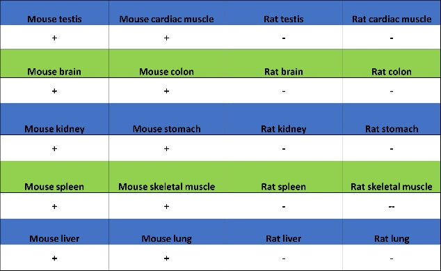
IHC shows positive staining in paraffin-embedded mouse cerebral cortex. Anti-Mouse TER-119/Erythroid Cells antibody was used at 1/500 dilution, followed by a HRP Polymer for Mouse & Rabbit IgG (ready to use). Counterstained with hematoxylin. Heat mediated antigen retrieval with Tris/EDTA buffer pH9.0 was performed before commencing with IHC staining protocol.
IHC shows positive staining in paraffin-embedded mouse liver. Anti-Mouse TER-119/Erythroid Cells antibody was used at 1/500 dilution, followed by a HRP Polymer for Mouse & Rabbit IgG (ready to use). Counterstained with hematoxylin. Heat mediated antigen retrieval with Tris/EDTA buffer pH9.0 was performed before commencing with IHC staining protocol.
IHC shows positive staining in paraffin-embedded mouse lung. Anti-Mouse TER-119/Erythroid Cells antibody was used at 1/500 dilution, followed by a HRP Polymer for Mouse & Rabbit IgG (ready to use). Counterstained with hematoxylin. Heat mediated antigen retrieval with Tris/EDTA buffer pH9.0 was performed before commencing with IHC staining protocol.