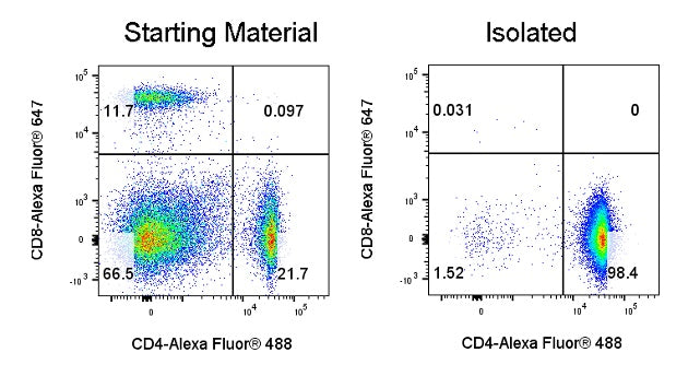
CD4+ T cells were enriched from mouse splenocytes using a column-based CD4+ T cell isolation kit. Cells were stained with CD8- Alexa Fluor® 647 (SDT Cat: S0B5895) and CD4- Alexa Fluor® 488 (SDT Cat: S0B8030) fluorescent antibodies, and analysis was performed by gating on live cells (Live+), allowing for the determination of the proportion of CD4+ T cells in the sorted fraction. Flow cytometry results indicated that the purity of CD4+ T cells after this sorting step was 98.4%.
Mouse CD4 T Cell Isolation Kit (Column-Based)
Mouse CD4 T Cell Isolation Kit (Column-Based)
Price:
Regular price
$767 USD
Regular price
Sale price
$767 USD
Unit price
per
For shipping services or bulk orders, you may request a quotation.
Secure checkout with

View full details
Product Details
Product Details
Product Specification
| Format |
1 mL Mouse CD4 T Cell Biotin-Antibody Cocktail 0.75mL Streptavidin Beads |
| Capacity | Suitable for a total cell count of 1×10⁹, and can perform up to 100 sorting runs. |
| Principle of Separation | This kit employs a column-based negative selection method to isolate CD4 T cells from mouse spleens. In this negative selection process, non-CD4 T cells are labeled with biotin-conjugated antibodies, and the cells are then incubated with streptavidin-nanomagnetic beads. As a result, the non-target cells become bound to the magnetic beads via the antibody‑bead complex, while the target CD4 T cells remain unlabeled. The cell suspension is subsequently loaded onto the separation column; under the influence of a magnetic field, the magnetically labeled non-target cells are retained within the column, whereas the unlabeled CD4 T cells elute by gravity into the collection tube. The resulting supernatant represents the CD4 T cell‑enriched fraction after removal of the non-target cells. |
| Reactivity | Mouse |
| Magnetic Bead Size | 20 nm |
| Separation Method | Column-Based |
| Selection Strategy | Negative Selection |
| Stability & Storage | Store protected from light at 2–8°C; do not freeze. |
Background
Mouse CD4 T lymphocytes are a core functional cell subset of the murine adaptive immune system. They originate from hematopoietic stem cells in the bone marrow, complete their development and maturation in the thymus, and then migrate to secondary lymphoid organs such as the spleen and lymph nodes. These cells specifically express the CD4 molecule on their surface and can recognize antigen peptides presented by antigen‑presenting cells (APCs) in complex with MHC class II molecules via the T‑cell receptor (TCR), thereby becoming activated and exerting immunoregulatory functions; they are often referred to as the "command cells" of the immune system. In scientific research, obtaining highly purified, high‑activity mouse CD4 T cells is essential for conducting related studies. This negative selection kit employs a negative sorting strategy: biotin‑conjugated antibodies label non‑CD4⁺ T cells, which are then bound by streptavidin‑coated magnetic nanoparticles and removed, keeping CD4 T cells untouched throughout the process. This approach maximizes the preservation of their native morphology and functional activity, providing reliable cellular material for subsequent experiments.
Protocol
Note: This kit does not include the sorting buffer; you need to prepare it yourself. The corresponding Stater catalog number isS0D3018
TakingL Separation Columnas an example
Steps |
Operating instructions |
Dosage and time |
|
|
Cell processing and labeling |
1 |
Take the mouse spleen, grind it on a 40 μm sterile filter mesh to prepare a single-cell suspension of the spleen, perform red blood cell lysis, and then count the cells. Note:It is recommended to count cells after staining with AO/PI; splenic cells can be centrifuged at 400 g for 7 minutes. |
Prepare the sample |
2 |
Resuspend 1×10^7 cells in 100 μL of MagSep Separation Buffer.7cells; Note:Adjust reagents proportionally according to the sample volume; set aside a pre-sorting sample to measure the initial CD4 ratio. |
1×10⁷cells/100 μL |
|
3 |
Add 10 μL of Mouse CD4 T cell Biotin Antibody Cocktail to the sample and gently mix. Note:In this step, gently pipette up and down 2–3 times to mix thoroughly; |
10 μL/100 μL |
|
4 |
Incubate the antibodies in the cocktail with the cells at room temperature for 10 min; |
Incubate at room temperature for 10 min |
|
5 |
Add MagSep Separation Buffer equal to ten times the cell volume to wash the cells, centrifuge at 400g for 7 min, discard the supernatant, and resuspend to the original volume; |
Centrifuge at 400g for 7 min |
|
6 |
Mix wellStreptavidin Beads, add 7.5 μLStreptavidin Beadsto the sample; |
7.5 μL/100 μL |
|
7 |
Gently mix the magnetic beads and cells, and incubate at room temperature for 5 min; |
Incubate at room temperature for 5 min |
|
Cell sorting |
8 |
In the column separation magnet, installL Separation Column, add 3 mL of MagSep Separation Buffer to rinse the separation column; |
Rinse the separation column |
9 |
Add the cells to the separation column and collect the target cells using a collection tube; Note:The cell volume should be at least 500 μL; if insufficient, make up the volume with MagSep Separation Buffer; the cells that drip down under gravity are the target cells; |
Collect the target cells |
|
10 |
Add 3 mL of MagSep Separation Buffer and collect the target cells; Note:This step elutes the unlabeled target cells from the separation column, increasing the yield. |
Collect the target cells |
|
|
Notes: 1.Pay attention to the maximum capacity of the separation column,The maximum number of labeled cells for the S Separation Column is1×107;The maximum number of labeled cells for the L Separation Column is1×108; 2.If the viability of the sample is low, it is recommended to use a dead cell removal kit to eliminate dead cells (Dead Cell Removal Set, (RUO)#S0K0009); | |||
Picture
Picture
Validation Data