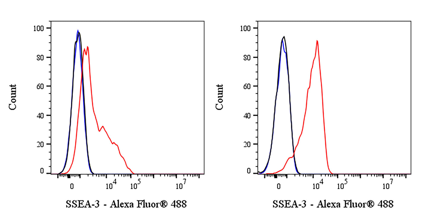
Flow cytometric analysis of HeLa (Human cervix adenocarcinoma epithelial cell, left) / NCCIT (Human pluripotent embryonic carcinoma epithelial cell, right) labeled SSEA-3 antibody at 1/400 dilution (0.5 μg) / (red) compared with a Mouse monoclonal IgG (black) Isotype Control and an unlabelled control (cells without incubation with primary antibody and secondary antibody) (blue). Goat Anti - Mouse IgM Alexa Fluor® 488 was used as the secondary antibody. Flow cytometry and data analysis were performed using Agilent NovoCyte Quanteon and FlowJo™ software.
Negative control: HeLa
Mouse Anti-Human SSEA-3 Antibody (S-3079)
Mouse Anti-Human SSEA-3 Antibody (S-3079)
Price:
Regular price
$100 USD
Regular price
Sale price
$100 USD
Unit price
per
For shipping services or bulk orders, you may request a quotation.
Secure checkout with

View full details
Product Details
Product Details
Product Specification
| Host | Mouse |
| Antigen | SSEA-3 |
| Location | Cell membrane |
| Clone Number | S-3079 |
| Antibody Type | Mouse mAb |
| Application | FCM |
| Reactivity | Hu |
| Positive Sample | NCCIT |
| Concentration | 2 mg/ml |
| Conjugation | Unconjugated |
| Physical Appearance | Liquid |
| Storage Buffer | PBS pH7.4 |
| Stability & Storage | 12 months from date of receipt / reconstitution, 2 to 8 °C as supplied |
Dilution
| application | dilution | species |
| FCM | 1:400 | Hu |
Background
SSEA-3 (Stage-Specific Embryonic Antigen-3) is a glycolipid surface antigen originally named for its specific expression during particular stages of mammalian embryonic development, and it is now widely used as a key marker for identifying the pluripotency of human embryonic stem cells and germ cells. In adult tissues, SSEA-3 is naturally present in the skin and bone marrow, serving as the core surface marker for "Muse cells" (Multilineage-differentiating Stress-Enduring cells); these cells possess tri-lineage differentiation potential without requiring genetic reprogramming, demonstrating immense promise in tissue repair and regenerative medicine. Furthermore, SSEA-3 holds significant pathological relevance, as its aberrant overexpression in certain malignancies (such as breast cancer) and cancer cell lines suggests a potential role in tumorigenesis and progression.
Picture
Picture
FC