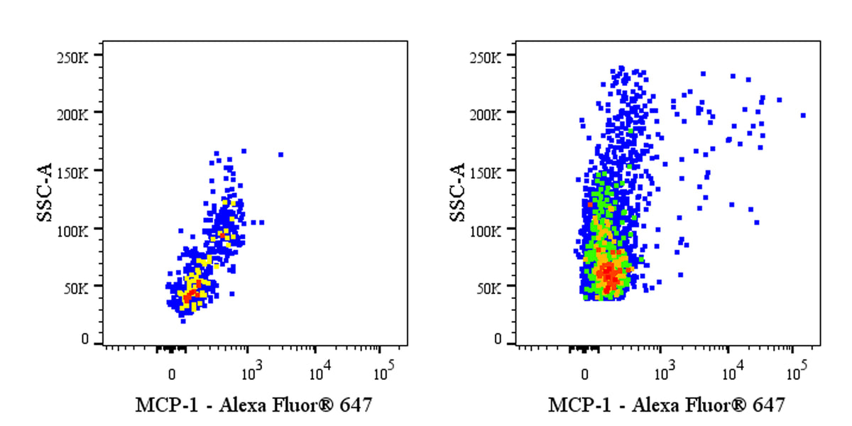
Flow cytometric analysis of human PBMCs (human peripheral blood mononuclear cells) stimulated with 10ng/ml lipopolysaccharide for 6hr in the presence of 1 μg/ml BFA (right panel) or untreated (left panel). The cells were harvested and fixed with 4% PFA and permeabilized with Intracellular Fixation & Permeabilization Buffer Set. The cells were then labeled with Anti-Human MCP-1 antibody at 1/200 dilution (1 μg). Goat Anti-Mouse IgG Alexa Fluor® 488 was used as the secondary antibody. Flow cytometry and data analysis were performed using BD FACSymphony™ A1 and FlowJo™ software.
Mouse Anti-Human MCP-1 Antibody (S-5583)
Mouse Anti-Human MCP-1 Antibody (S-5583)
Price:
Regular price
$100 USD
Regular price
Sale price
$100 USD
Unit price
per
For shipping services or bulk orders, you may request a quotation.
Secure checkout with

View full details
Product Details
Product Details
Product Specification
| Host | Mouse |
| Antigen | CCL2 |
| Location | Secreted |
| Accession | P13500 |
| Clone Number | S-5583 |
| Antibody Type | Mouse mAb |
| Isotype Control | S0B0617 |
| Application | ICFCM |
| Reactivity | Hu |
| Positive Sample | Human PBMCs stimulated with LPS and BFA |
| Purification | Protein A |
| Concentration | 2 mg/ml |
| Conjugation | Unconjugated |
| Physical Appearance | Liquid |
| Storage Buffer | PBS pH7.4 |
| Stability & Storage | 12 months from date of receipt / reconstitution, 2 to 8 °C as supplied |
Dilution
| application | dilution | species |
| ICFCM | 1:200 | Hu |
Background
Monocyte chemoattractant protein-1 (MCP-1), also known as C-C motif chemokine ligand 2 (CCL2), is a 76-amino-acid glycoprotein of approximately 8.6 kDa belonging to the CC chemokine family, encoded by the CCL2 gene on human chromosome 17q12. Its structure features four conserved cysteine residues forming two intramolecular disulfide bonds, with the N-terminal residues 1–6 essential for chemoattractant activity and residue 14 subject to glycosylation. MCP-1 is secreted by a wide variety of cells including monocytes, fibroblasts, endothelial cells, epithelial cells, smooth muscle cells, and various tumor cells in response to stimuli such as TNF-α, IFN-γ, and PDGF. Functionally, MCP-1 is a potent chemoattractant that binds its primary receptor CCR2 on monocytes, triggering chemotaxis, firm adhesion to vascular endothelium, and transendothelial migration to sites of inflammation; it also attracts memory T lymphocytes and natural killer cells. MCP-1 plays a critical role in numerous inflammatory pathologies including atherosclerosis, rheumatoid arthritis, renal disease, and tumor development, where it promotes macrophage infiltration that exacerbates disease progression. Beyond inflammation, emerging evidence implicates MCP-1 in Alzheimer's disease pathogenesis, where it participates in neuroinflammation and amyloid-β deposition, positioning it as a potential early diagnostic biomarker and therapeutic target.
Picture
Picture
FC