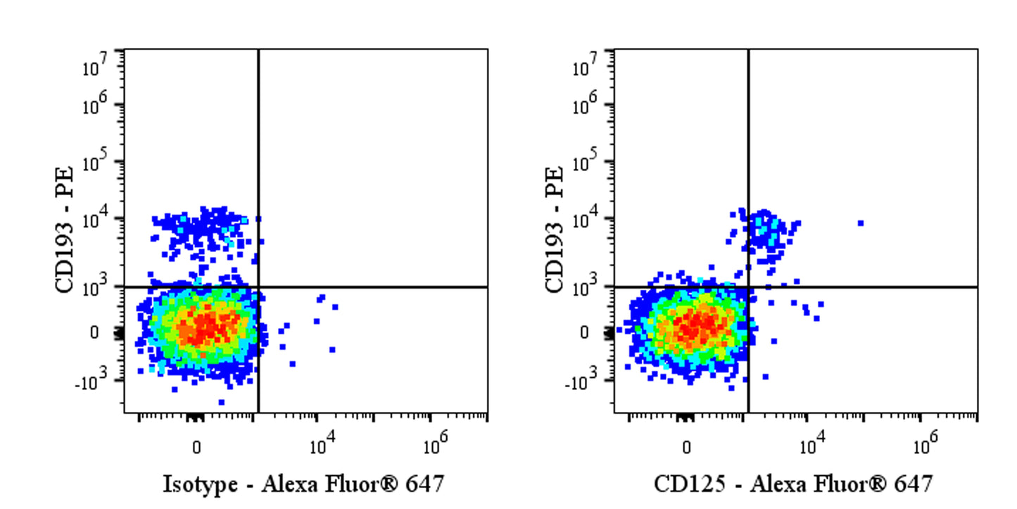
Flow cytometric analysis of human peripheral blood cells labeled with Human CD125 antibody at 1/200 dilution (1 μg) / (right panel) compared with a Mouse IgG1, κ Isotype Control / (left panel). Goat Anti-Mouse IgG Alexa Fluor® 647 was used as the secondary antibody. Then cells were stained with CD193 – PE antibody separately. Total viable cells, as determined by Fixable Viability Dye 545 (S0B88802), were used for analysis. Flow cytometry and data analysis were performed using Agilent NovoCyte Quanteon and FlowJo™ software.
Mouse Anti-Human CD125 Antibody (S-4480)
Mouse Anti-Human CD125 Antibody (S-4480)
Price:
Regular price
$100 USD
Regular price
Sale price
$100 USD
Unit price
per
For shipping services or bulk orders, you may request a quotation.
Secure checkout with

View full details
Product Details
Product Details
Product Specification
| Host | Mouse |
| Antigen | CD125 |
| Synonyms | Interleukin-5 receptor subunit alpha; IL-5 receptor subunit alpha; IL-5R subunit alpha; IL-5R-alpha; IL-5RAAlternative nameCDw125; IL5R; IL5RA |
| Location | Membrane |
| Accession | Q01344 |
| Clone Number | S-4480 |
| Antibody Type | Mouse mAb |
| Isotype | IgG1,k |
| Application | FCM |
| Reactivity | Hu |
| Positive Sample | Human peripheral blood cells |
| Purification | Protein G |
| Concentration | 2 mg/ml |
| Conjugation | Unconjugated |
| Physical Appearance | Liquid |
| Storage Buffer | PBS pH7.4 |
| Stability & Storage | 12 months from date of receipt / reconstitution, 2 to 8 °C as supplied |
Dilution
| application | dilution | species |
| FCM | 1:200 | Hu |
Background
CD125, also known as the interleukin-4 receptor subunit alpha (IL-4Rα), is a type I transmembrane protein that serves as a critical shared signaling component for both the type I and type II interleukin-4 receptors, thereby playing a pivotal role in regulating immune responses, particularly those associated with Th2-mediated inflammation, allergic reactions, and asthma. As the primary binding subunit for interleukin-4 (IL-4) and interleukin-13 (IL-13), CD125 facilitates the activation of downstream signaling pathways, most notably the JAK-STAT pathway (specifically JAK1/STAT6), which leads to the transcription of genes involved in IgE class switching, eosinophil recruitment, mucus production, and alternative macrophage activation. Structurally, it consists of an extracellular domain responsible for cytokine binding, a single transmembrane helix, and a cytoplasmic tail containing specific motifs that recruit Janus kinases, making it a significant therapeutic target; indeed, monoclonal antibodies such as dupilumab specifically inhibit the interaction of IL-4 and IL-13 with CD125 to effectively treat moderate-to-severe atopic dermatitis, asthma, and chronic rhinosinusitis with nasal polyps by blocking this central axis of type 2 inflammatory signaling.
Picture
Picture
FC