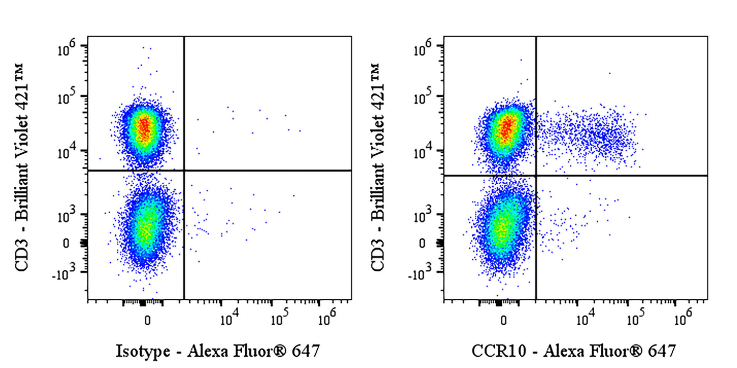
Flow cytometric analysis of human PBMC labeled with anti-CCR10 antibody at 1/2000 dilution (0.1 μg) / (right panel) compared with a Mouse IgG2a, κ Isotype Control / (left panel). Goat Anti-Mouse IgG Alexa Fluor® 647 was used as the secondary antibody. Then cells were stained with CD3 - Brilliant Violet 421™ antibody separately. Flow cytometry and data analysis were performed using Agilent NovoCyte Quanteon and FlowJo™ software.
Mouse Anti-Human CCR10 Antibody (S-4710)
Mouse Anti-Human CCR10 Antibody (S-4710)
Price:
Regular price
$100 USD
Regular price
Sale price
$100 USD
Unit price
per
For shipping services or bulk orders, you may request a quotation.
Secure checkout with

View full details
Product Details
Product Details
Product Specification
| Host | Mouse |
| Antigen | CCR10 |
| Synonyms | C-C chemokine receptor type 10; C-C CKR-10; CC-CKR-10; CCR-10Alternative nameG-protein coupled receptor 2; GPR2 |
| Location | Cell membrane |
| Accession | P46092 |
| Clone Number | S-4710 |
| Antibody Type | Mouse mAb |
| Isotype | IgG2a,k |
| Application | FCM |
| Reactivity | Hu |
| Positive Sample | Human PBMC |
| Purification | Protein A |
| Concentration | 2 mg/ml |
| Conjugation | Unconjugated |
| Physical Appearance | Liquid |
| Storage Buffer | PBS pH7.4 |
| Stability & Storage | 12 months from date of receipt / reconstitution, 2 to 8 °C as supplied |
Dilution
| application | dilution | species |
| FCM | 1:2000 | Hu |
Background
The CCR10 protein (C-C motif chemokine receptor 10) is a G protein-coupled receptor primarily expressed on the surface of skin T cells, IgA plasmablasts, and various skin structural cells (such as fibroblasts and melanocytes). It primarily regulates the specific migration (homing) of lymphocytes to the skin and mucosal tissues such as the intestine and lungs by binding to its ligands, CCL27 (highly expressed in keratinocytes) and CCL28 (highly expressed in mucosal epithelial cells). This receptor plays a critical role in maintaining skin immune homeostasis, promoting wound healing, and mediating the localization of IgA-secreting cells to mucosal sites such as the mammary gland. However, its function is highly context-dependent: in inflammatory diseases such as psoriasis and atopic dermatitis, CCR10 can both mediate the skin infiltration of pathogenic T cells and help limit excessive inflammatory responses. In cancers such as melanoma, it may also be exploited by tumor cells to achieve immune evasion and metastasis.
Picture
Picture
FC