WB result of Monoamine Oxidase B/MAOB Recombinant Rabbit mAb
Primary antibody: Monoamine Oxidase B/MAOB Recombinant Rabbit mAb at 1/1000 dilution
Lane 1: A204 whole cell lysate 20 µg
Lane 2: HepG2 whole cell lysate 20 µg
Lane 3: MCF7 whole cell lysate 20 µg
Lane 4: SH-SY5Y whole cell lysate 20 µg
Negative control: A204 whole cell lysate
Secondary antibody: Goat Anti- rabbit IgG, (H+L), HRP conjugated at 1/10000 dilution
Predicted MW: 59 kDa
Observed MW: 60 kDa
This blot was developed with high sensitivity substrate
Monoamine Oxidase B/MAOB Recombinant Rabbit mAb (S-5317)
Monoamine Oxidase B/MAOB Recombinant Rabbit mAb (S-5317)
Price:
Regular price
$100 USD
Regular price
Sale price
$100 USD
Unit price
per
For shipping services or bulk orders, you may request a quotation.
Secure checkout with

View full details
Product Details
Product Details
Product Specification
| Host | Rabbit |
| Antigen | Monoamine Oxidase B/MAOB |
| Synonyms | Amine oxidase [flavin-containing] B |
| Location | Mitochondrion |
| Accession | P27338 |
| Clone Number | S-5317 |
| Antibody Type | Recombinant mAb |
| Isotype | IgG |
| Application | WB, IHC-P |
| Reactivity | Hu |
| Positive Sample | HepG2, MCF7, SH-SY5Y |
| Purification | Protein A |
| Concentration | 0.5 mg/ml |
| Conjugation | Unconjugated |
| Physical Appearance | Liquid |
| Storage Buffer | PBS, 40% Glycerol, 0.05% BSA, 0.02% sodium azide |
| Stability & Storage | 12 months from date of receipt / reconstitution, -20 °C as supplied |
Dilution
| application | dilution | species |
| WB | 1:500-1:1000 | Hu |
| IHC-P | 1:250 | Hu |
Background
Monoamine oxidase B (MAO-B) is an enzyme primarily found in the brain and platelets that catalyzes the oxidative deamination of neuroactive and vasoactive amines, particularly dopamine. By breaking down dopamine, MAO-B plays a key role in regulating its synaptic levels, making it an important pharmacological target—MAO-B inhibitors (e.g., selegiline, rasagiline) are used as adjunct therapies in Parkinson’s disease to enhance dopaminergic neurotransmission and provide neuroprotective effects.
Picture
Picture
Western Blot
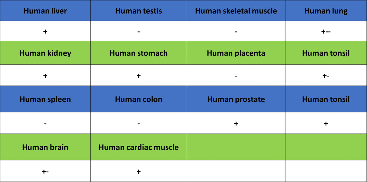
Immunohistochemistry
IHC shows positive staining in paraffin-embedded human cerebral cortex. Anti-MAOB antibody was used at 1/250 dilution, followed by a HRP Polymer for Rabbit IgG (ready to use). Counterstained with hematoxylin. Heat mediated antigen retrieval with Tris/EDTA buffer pH9.0 was performed before commencing with IHC staining protocol.
IHC shows positive staining in paraffin-embedded human cardiac muscle. Anti-MAOB antibody was used at 1/250 dilution, followed by a HRP Polymer for Rabbit IgG (ready to use). Counterstained with hematoxylin. Heat mediated antigen retrieval with Tris/EDTA buffer pH9.0 was performed before commencing with IHC staining protocol.
IHC shows positive staining in paraffin-embedded human kidney. Anti-MAOB antibody was used at 1/250 dilution, followed by a HRP Polymer for Rabbit IgG (ready to use). Counterstained with hematoxylin. Heat mediated antigen retrieval with Tris/EDTA buffer pH9.0 was performed before commencing with IHC staining protocol.
IHC shows positive staining in paraffin-embedded human liver. Anti-MAOB antibody was used at 1/250 dilution, followed by a HRP Polymer for Rabbit IgG (ready to use). Counterstained with hematoxylin. Heat mediated antigen retrieval with Tris/EDTA buffer pH9.0 was performed before commencing with IHC staining protocol.
IHC shows positive staining in paraffin-embedded human pancreatic cancer. Anti-MAOB antibody was used at 1/250 dilution, followed by a HRP Polymer for Rabbit IgG (ready to use). Counterstained with hematoxylin. Heat mediated antigen retrieval with Tris/EDTA buffer pH9.0 was performed before commencing with IHC staining protocol.