Standard curve
Example of IL-1β standard curve in Assay Diluent .
Monkey IL-1β Single Plex Assay (适用于Luminex)
Monkey IL-1β Single Plex Assay (适用于Luminex)
Price:
Regular price
$585 USD
Regular price
Sale price
$585 USD
Unit price
per
For shipping services or bulk orders, you may request a quotation.
Secure checkout with

View full details
Product Details
Product Details
Product Specification
| Antigen | IL-1β |
| Reactivity | Monkey |
| Stability & Storage | 12 months from date of receipt / reconstitution, 2 to 8°C as supplied. |
Kit
| Precision | Intra-assay: <10%; Inter-assay: <15% |
| Sample type | Serum; Plasma; Cell culture supernatant |
| Assay type | Sandwich (quantitative) |
| Sensitivity | <1 pg/mL |
| Range | 1.08pg/mL-100000pg/mL |
| Recovery | Plasma: 80%-120% Serum: 80%-120% Cell culture supernatant: 80%-120% |
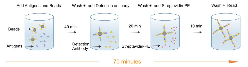
| Assay time | 70 minutes |
| Species reactivity | Monkey |
Background
Monkey IL-1β (interleukin-1 beta) is a recombinant cytokine derived from non-human primates such as cynomolgus or rhesus macaques. As a key mediator of innate immunity, it plays a central role in promoting inflammatory responses to infection, tissue damage, and autoimmune stimuli. Due to its high structural and functional homology with human IL-1β, monkey IL-1β serves as a critical tool in translational research. It is widely used in immune cell activation assays, inflammasome studies, and preclinical evaluation of anti-inflammatory biologics. The protein’s strong cross-reactivity with human IL-1 receptors enables accurate modeling of human inflammatory diseases in primate systems, providing essential insights into drug efficacy and safety prior to clinical trials.
Picture
Picture
Luminex
Quantifiction of Monkey PBMC
Stimulated sample: Monkey peripheral blood mononuclear cells (PBMCs) were stimulated with PHA、LPS、Lono and the average concentration of IL-1β in the supernatant was measured to be 106.23 pg/mL.
Unstimulated sample: Monkey PBMCs were cultured for 24h, and the average concentration of IL-1β in the supernatant was measured to be 7.47 pg/mL.
Recovery
Within the detection range, the combined cytokine proteins were added to serum,plasma,cell supernatant at high、low concentrations.The matrices used in these experiments were diluted 2-fold with the assay buffer before the addition of the cytokine proteins. The results were compared with those of samples that had the same concentration of cytokines added in the assay buffer.
Protocol Diagram