WB result of MOG Rabbit Polyclonal Antibody
Primary antibody: MOG Rabbit Polyclonal Antibody at 1/1000 dilution
Lane 1: mouse lung lysate 20 µg
Lane 2: mouse brain lysate 20 µg
Negative control: mouse lung lysate
Secondary antibody: Goat Anti-Rabbit IgG (H+L), HRP conjugated at 1/10000 dilution
Predicted MW: 28 kDa
Observed MW: 24 kDa
MOG Rabbit Polyclonal Antibody
MOG Rabbit Polyclonal Antibody
Price:
Regular price
$100 USD
Regular price
Sale price
$100 USD
Unit price
per
For shipping services or bulk orders, you may request a quotation.
Secure checkout with

View full details
Product Details
Product Details
Product Specification
| Host | Rabbit |
| Antigen | MOG |
| Synonyms | Myelin-oligodendrocyte glycoprotein |
| Immunogen | Recombinant Protein |
| Location | Cell membrane |
| Accession | Q16653 |
| Antibody Type | Polyclonal antibody |
| Isotype | IgG |
| Application | WB, IHC-P, IF |
| Reactivity | Hu, Ms, Rt |
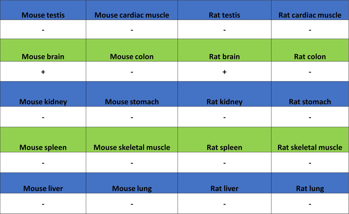
| Positive Sample | mouse brain, rat brain |
| Purification | Immunogen Affinity |
| Concentration | 0.5 mg/ml |
| Conjugation | Unconjugated |
| Physical Appearance | Liquid |
| Storage Buffer | PBS, 40% Glycerol, 0.05% BSA, 0.02% sodium azide |
| Stability & Storage | 12 months from date of receipt / reconstitution, -20 °C as supplied |
Dilution
| application | dilution | species |
| WB | 1:1000-1:5000 | Ms, Rt |
| IHC-P | 1:200-1:1000 | Hu, Ms, Rt |
| IF | 1:1000 | Hu, Ms, Rt |
Background
MOG (Myelin Oligodendrocyte Glycoprotein) is a type I integral membrane protein expressed exclusively in the central nervous system, specifically localized to the outermost layer of myelin sheaths and the surface membrane of oligodendrocytes. Its extracellular domain adopts an immunoglobulin-like (IgV) fold and can form antiparallel homodimers, suggesting that it may play a physiological role in mediating adhesion between myelin membranes or in maintaining the structural integrity of myelin sheaths. However, the core reason MOG protein draws intense medical attention is that it serves as a critical autoantigen in inflammatory demyelinating diseases of the central nervous system: when immune tolerance is disrupted, the generated anti-MOG autoantibodies (MOG-IgG) can target this protein, giving rise to a group of recently formally defined independent disease entities—MOG antibody-associated disease (MOGAD). This disease presents with diverse clinical manifestations, predominantly affects children and young adults, and commonly features optic neuritis, acute disseminated encephalomyelitis, myelitis, and brainstem encephalitis, with approximately 50%–60% of patients following a relapsing course. In terms of diagnosis, the cell-based assay (CBA) using full-length human MOG is the gold standard for detecting MOG-IgG, with serum antibody titers often correlating with disease activity and potentially turning negative after treatment; in terms of prognosis, MOGAD is generally considered distinct from multiple sclerosis or aquaporin-4 antibody-positive neuromyelitis optica spectrum disorders, shows good response to corticosteroid therapy, but repeated attacks may still lead to neurological disability.
Picture
Picture
Western Blot
WB result of MOG Rabbit Polyclonal Antibody
Primary antibody: MOG Rabbit Polyclonal Antibody at 1/1000 dilution
Lane 1: rat lung lysate 20 µg
Lane 2: rat brain lysate 20 µg
Negative control: rat lung lysate
Secondary antibody: Goat Anti-Rabbit IgG (H+L), HRP conjugated at 1/10000 dilution
Predicted MW: 28 kDa
Observed MW: 24 kDa
Immunohistochemistry
IHC shows positive staining in paraffin-embedded human cerebral cortex. Anti-MOG antibody was used at 1/1000 dilution, followed by a HRP Polymer for Mouse & Rabbit IgG (ready to use). Counterstained with hematoxylin. Heat mediated antigen retrieval with Tris/EDTA buffer pH9.0 was performed before commencing with IHC staining protocol.
Negative control: IHC shows negative staining in paraffin-embedded human colon. Anti-MOG antibody was used at 1/1000 dilution, followed by a HRP Polymer for Mouse & Rabbit IgG (ready to use). Counterstained with hematoxylin. Heat mediated antigen retrieval with Tris/EDTA buffer pH9.0 was performed before commencing with IHC staining protocol.
IHC shows positive staining in paraffin-embedded mouse cerebral cortex. Anti-MOG antibody was used at 1/1000 dilution, followed by a HRP Polymer for Mouse & Rabbit IgG (ready to use). Counterstained with hematoxylin. Heat mediated antigen retrieval with Tris/EDTA buffer pH9.0 was performed before commencing with IHC staining protocol.
Negative control: IHC shows negative staining in paraffin-embedded mouse colon. Anti-MOG antibody was used at 1/1000 dilution, followed by a HRP Polymer for Mouse & Rabbit IgG (ready to use). Counterstained with hematoxylin. Heat mediated antigen retrieval with Tris/EDTA buffer pH9.0 was performed before commencing with IHC staining protocol.
IHC shows positive staining in paraffin-embedded rat cerebral cortex. Anti-MOG antibody was used at 1/1000 dilution, followed by a HRP Polymer for Mouse & Rabbit IgG (ready to use). Counterstained with hematoxylin. Heat mediated antigen retrieval with Tris/EDTA buffer pH9.0 was performed before commencing with IHC staining protocol.
Negative control: IHC shows negative staining in paraffin-embedded rat colon. Anti-MOG antibody was used at 1/1000 dilution, followed by a HRP Polymer for Mouse & Rabbit IgG (ready to use). Counterstained with hematoxylin. Heat mediated antigen retrieval with Tris/EDTA buffer pH9.0 was performed before commencing with IHC staining protocol.
Immunofluorescence
IF shows positive staining in paraffin-embedded human cerebral cortex. Anti-MOG antibody was used at 1/1000 dilution (Green) and incubated overnight at 4°C. Goat polyclonal Antibody to Rabbit IgG - H&L (Alexa Fluor® 488) was used as secondary antibody at 1/1000 dilution. Counterstained with DAPI (Blue). Heat mediated antigen retrieval with EDTA buffer pH9.0 was performed before commencing with IF staining protocol.
IF shows positive staining in paraffin-embedded mouse cerebral cortex. Anti-MOG antibody was used at 1/1000 dilution (Green) and incubated overnight at 4°C. Goat polyclonal Antibody to Rabbit IgG - H&L (Alexa Fluor® 488) was used as secondary antibody at 1/1000 dilution. Counterstained with DAPI (Blue). Heat mediated antigen retrieval with EDTA buffer pH9.0 was performed before commencing with IF staining protocol.
IF shows positive staining in paraffin-embedded rat cerebral cortex. Anti-MOG antibody was used at 1/1000 dilution (Green) and incubated overnight at 4°C. Goat polyclonal Antibody to Rabbit IgG - H&L (Alexa Fluor® 488) was used as secondary antibody at 1/1000 dilution. Counterstained with DAPI (Blue). Heat mediated antigen retrieval with EDTA buffer pH9.0 was performed before commencing with IF staining protocol.