WB result of Moesin Rabbit Polyclonal Antibody
Primary antibody: Moesin Rabbit Polyclonal Antibody at 1/1000 dilution
Lane 1: HeLa whole cell lysate 20 μg
Lane 2: Raji whole cell lysate 20 μg
Lane 3: Jurkat whole cell lysate 20 μg
Lane 4: HUVEC whole cell lysate 20 μg
Secondary antibody: Goat Anti-Rabbit IgG (H+L), HRP conjugated at 1/10000 dilution
Predicted MW: 68 kDa
Observed MW: 78 kDa
Moesin Rabbit Polyclonal Antibody
Moesin Rabbit Polyclonal Antibody
Price:
Regular price
$100 USD
Regular price
Sale price
$100 USD
Unit price
per
For shipping services or bulk orders, you may request a quotation.
Secure checkout with

View full details
Product Details
Product Details
Product Specification
| Host | Rabbit |
| Antigen | Moesin |
| Immunogen | Synthetic Peptide |
| Location | Cell membrane |
| Accession | P26038 |
| Antibody Type | Polyclonal antibody |
| Isotype | IgG |
| Application | WB, IHC-P, IF |
| Reactivity | Hu, Ms, Rt |
| Positive Sample | HeLa, Raji, Jurkat, HUVEC, NIH/3T3, mouse spleen, C6, mouse spleen |
| Predicted Reactivity | Bv |
| Purification | Immunogen Affinity |
| Concentration | 0.5 mg/ml |
| Conjugation | Unconjugated |
| Physical Appearance | Liquid |
| Storage Buffer | PBS, 40% Glycerol, 0.05% BSA, 0.02% sodium azide |
| Stability & Storage | 12 months from date of receipt / reconstitution, -20 °C as supplied |
Dilution
| application | dilution | species |
| WB | 1:1000 | Hu, Ms, Rt |
| IHC-P | 1:100-1:250 | Hu, Ms, Rt |
| IF | 1:100 | Ms |
Background
Moesin (membrane-organizing extension spike protein) is a member of the ERM (ezrin-radixin-moesin) family, encoded by the MSN gene located on the X chromosome, comprising 577 amino acids with a molecular weight of approximately 68 kDa. As a cytoskeletal linker protein, Moesin binds to the plasma membrane or transmembrane proteins through its N-terminal FERM domain and anchors F-actin via its C-terminal actin-binding domain, thereby establishing a regulated physical connection between the plasma membrane and the cytoskeleton. In the resting state, the protein exists in an autoinhibited closed conformation in which the N- and C-termini mask each other's binding sites through head-to-tail interaction; upon phosphorylation of its C-terminal threonine or binding to phosphatidylinositol 4,5-bisphosphate (PIP2), the conformation opens and the protein becomes activated. Activated Moesin participates in the regulation of cell morphology, membrane trafficking, immune synapse formation, and lymphocyte migration, and is highly expressed in endothelial cells and leukocytes. Recent studies have further revealed the role of Moesin in disease: in glioblastoma, Moesin interacts with CD44 and promotes tumor proliferation and stemness maintenance through the Wnt/β-catenin pathway, correlating with poor prognosis; in the field of bone biology, Moesin regulates osteoclast fusion and bone resorption; in addition, Moesin is involved in HMGB1-mediated vascular hyperpermeability and septic inflammatory responses.
Picture
Picture
Western Blot
WB result of Moesin Rabbit Polyclonal Antibody
Primary antibody: Moesin Rabbit Polyclonal Antibody at 1/1000 dilution
Lane 1: NIH/3T3 whole cell lysate 20 μg
Lane 2: mouse spleen lysate 20 μg
Secondary antibody: Goat Anti-Rabbit IgG (H+L), HRP conjugated at 1/10000 dilution
Predicted MW: 68 kDa
Observed MW: 78 kDa
Primary antibody: Moesin Rabbit Polyclonal Antibody at 1/1000 dilution
Lane 1: C6 whole cell lysate 20 μg
Lane 2: rat spleen lysate 20 μg
Secondary antibody: Goat Anti-Rabbit IgG (H+L), HRP conjugated at 1/10000 dilution
Predicted MW: 68 kDa
Observed MW: 78 kDa
Immunohistochemistry
IHC shows positive staining in paraffin-embedded human spleen. Anti-Moesin antibody was used at 1/250 dilution, followed by a HRP Polymer for Rabbit IgG (ready to use). Counterstained with hematoxylin. Heat mediated antigen retrieval with Tris/EDTA buffer pH9.0 was performed before commencing with IHC staining protocol.
IHC shows positive staining in paraffin-embedded human bladder cancer. Anti-Moesin antibody was used at 1/250 dilution, followed by a HRP Polymer for Rabbit IgG (ready to use). Counterstained with hematoxylin. Heat mediated antigen retrieval with Tris/EDTA buffer pH9.0 was performed before commencing with IHC staining protocol.
IHC shows positive staining in paraffin-embedded human colon cancer. Anti-Moesin antibody was used at 1/250 dilution, followed by a HRP Polymer for Rabbit IgG (ready to use). Counterstained with hematoxylin. Heat mediated antigen retrieval with Tris/EDTA buffer pH9.0 was performed before commencing with IHC staining protocol.
IHC shows positive staining in paraffin-embedded mouse spleen. Anti-Moesin antibody was used at 1/250 dilution, followed by a HRP Polymer for Rabbit IgG (ready to use). Counterstained with hematoxylin. Heat mediated antigen retrieval with Tris/EDTA buffer pH9.0 was performed before commencing with IHC staining protocol.
IHC shows positive staining in paraffin-embedded rat spleen. Anti-Moesin antibody was used at 1/250 dilution, followed by a HRP Polymer for Rabbit IgG (ready to use). Counterstained with hematoxylin. Heat mediated antigen retrieval with Tris/EDTA buffer pH9.0 was performed before commencing with IHC staining protocol.
Immunofluorescence
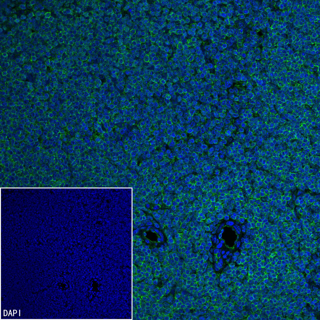
IF shows positive staining in paraffin-embedded mouse spleen. Anti-Moesin antibody was used at 1/100 dilution (Green) and incubated overnight at 4°C. Goat polyclonal Antibody to Rabbit IgG - H&L (Alexa Fluor® 488) was used as secondary antibody at 1/1000 dilution. Counterstained with DAPI (Blue). Heat mediated antigen retrieval with EDTA buffer pH9.0 was performed before commencing with IF staining protocol.