WB result of MMP-8 Recombinant Rabbit mAb
Primary antibody: MMP-8 Recombinant Rabbit mAb at 1/1000 dilution
Lane 1: mouse spleen lysate 20 µg
Lane 2: mouse lung lysate 20 µg
Secondary antibody: Goat Anti-Rabbit IgG (H+L), HRP conjugated at 1/10000 dilution
Predicted MW: 53 kDa
Observed MW: 60 kDa
MMP-8 Recombinant Rabbit mAb (S-5674)
MMP-8 Recombinant Rabbit mAb (S-5674)
Price:
Regular price
$100 USD
Regular price
Sale price
$100 USD
Unit price
per
For shipping services or bulk orders, you may request a quotation.
Secure checkout with

View full details
Product Details
Product Details
Product Specification
| Host | Rabbit |
| Antigen | MMP-8 |
| Synonyms | Neutrophil collagenase; Matrix metalloproteinase-8 (MMP-8); PMNL collagenase (PMNL-CL); CLG1; MMP8 |
| Location | Secreted |
| Accession | P22894 |
| Clone Number | S-5674 |
| Antibody Type | Recombinant mAb |
| Isotype | IgG |
| Application | WB, IHC-P |
| Reactivity | Hu, Ms, Rt |
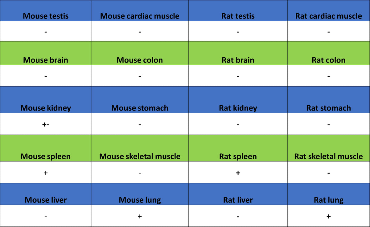
| Positive Sample | mouse spleen, mouse lung, rat lung |
| Purification | Protein A |
| Concentration | 2 mg/ml |
| Conjugation | Unconjugated |
| Physical Appearance | Liquid |
| Storage Buffer | PBS, 40% Glycerol, 0.05% BSA, 0.02% sodium azide |
| Stability & Storage | 12 months from date of receipt / reconstitution, -20 °C as supplied |
Dilution
| application | dilution | species |
| WB | 1:1000 | Ms, Rt |
| IHC-P | 1:2000 | Hu, Ms, Rt |
Background
MMP-8, also known as neutrophil collagenase, is a zinc-dependent metalloproteinase encoded by the MMP8 gene. It is primarily expressed in neutrophils and is one of the key enzymes that initiates the degradation of fibrillar collagens, particularly type I collagen. In terms of physiological function, MMP-8 is not only involved in neural crest cell migration and tissue remodeling during embryonic development, but also plays a significant role in pathological processes such as atherosclerosis due to its potent collagenolytic activity—it degrades collagen in the fibrous cap of plaques, which is associated with plaque instability and increased risk of cardiovascular disease. In cancer research, the role of MMP-8 is complex, exhibiting both tumor-suppressive and tumor-promoting properties depending on the tumor type and microenvironment. As one of the most potent interstitial collagenases in degrading collagen, MMP-8 activity is finely regulated in vivo, making it a key participant in various inflammatory diseases and matrix remodeling-related disorders, as well as a potential therapeutic target.
Picture
Picture
Western Blot
WB result of MMP-8 Recombinant Rabbit mAb
Primary antibody: MMP-8 Recombinant Rabbit mAb at 1/1000 dilution
Lane 1: rat lung lysate 20 µg
Secondary antibody: Goat Anti-Rabbit IgG (H+L), HRP conjugated at 1/10000 dilution
Predicted MW: 53 kDa
Observed MW: 60 kDa
Immunohistochemistry
IHC shows positive staining in paraffin-embedded human spleen. Anti-MMP-8 antibody was used at 1/2000 dilution, followed by a HRP Polymer for Rabbit IgG (ready to use). Counterstained with hematoxylin. Heat mediated antigen retrieval with Tris/EDTA buffer pH9.0 was performed before commencing with IHC staining protocol.
IHC shows positive staining in paraffin-embedded human colon cancer. Anti-MMP-8 antibody was used at 1/2000 dilution, followed by a HRP Polymer for Rabbit IgG (ready to use). Counterstained with hematoxylin. Heat mediated antigen retrieval with Tris/EDTA buffer pH9.0 was performed before commencing with IHC staining protocol.
IHC shows positive staining in paraffin-embedded human gastric cancer. Anti-MMP-8 antibody was used at 1/2000 dilution, followed by a HRP Polymer for Rabbit IgG (ready to use). Counterstained with hematoxylin. Heat mediated antigen retrieval with Tris/EDTA buffer pH9.0 was performed before commencing with IHC staining protocol.
IHC shows positive staining in paraffin-embedded mouse spleen. Anti-MMP-8 antibody was used at 1/2000 dilution, followed by a HRP Polymer for Rabbit IgG (ready to use). Counterstained with hematoxylin. Heat mediated antigen retrieval with Tris/EDTA buffer pH9.0 was performed before commencing with IHC staining protocol.
IHC shows positive staining in paraffin-embedded rat spleen. Anti-MMP-8 antibody was used at 1/2000 dilution, followed by a HRP Polymer for Rabbit IgG (ready to use). Counterstained with hematoxylin. Heat mediated antigen retrieval with Tris/EDTA buffer pH9.0 was performed before commencing with IHC staining protocol.