WB result of MEF2B Recombinant Rabbit mAb
Primary antibody: MEF2B Recombinant Rabbit mAb at 1/1000 dilution
Lane 1: HeLa whole cell lysate 20 µg
Lane 2: Raji whole cell lysate 20 µg
Lane 3: Daudi whole cell lysate 20 µg
Lane 4: Ramos whole cell lysate 20 µg
Negative control: HeLa whole cell lysate
Low expression control: Raji whole cell lysate
Secondary antibody: Goat Anti- rabbit IgG, (H+L), HRP conjugated at 1/10000 dilution
Predicted MW: 38 kDa
Observed MW: 40 kDa
MEF2B Recombinant Rabbit mAb (S-3788-28)
MEF2B Recombinant Rabbit mAb (S-3788-28)
Price:
Regular price
$100 USD
Regular price
Sale price
$100 USD
Unit price
per
For shipping services or bulk orders, you may request a quotation.
Secure checkout with

View full details
Product Details
Product Details
Product Specification
| Host | Rabbit |
| Antigen | MEF2B |
| Synonyms | Myocyte-specific enhancer factor 2B; RSRFR2; Serum response factor-like protein 2; XMEF2 |
| Immunogen | Recombinant Protein |
| Location | Nucleus |
| Accession | Q02080 |
| Clone Number | S-3788-28 |
| Antibody Type | Recombinant mAb |
| Isotype | IgG |
| Application | WB, IHC-P, ICC |
| Reactivity | Hu |
| Positive Sample | Raji, Daudi, Ramos |
| Purification | Protein A |
| Concentration | 0.5 mg/ml |
| Conjugation | Unconjugated |
| Physical Appearance | Liquid |
| Storage Buffer | PBS, 40% Glycerol, 0.05% BSA, 0.03% Proclin 300 |
| Stability & Storage | 12 months from date of receipt / reconstitution, -20 °C as supplied |
Dilution
| application | dilution | species |
| WB | 1:1000 | Hu |
| IHC-P | 1:200 | Hu |
| ICC | 1:500 | Hu |
Background
The MEF2B protein is a member of the myocyte enhancer factor 2 (MEF2) transcription factor family, highly expressed primarily in the nervous and lymphatic systems, and plays a critical role in regulating gene transcription. Unlike other MEF2 family members (such as MEF2A and MEF2D), MEF2B exhibits unique tissue distribution and functional specificity. It binds to specific DNA sequences (A/T-rich elements) via its conserved MADS box and MEF2 domains, and requires interactions with cofactors (such as MyoD and MAPK signaling pathway-regulated co-activators or co-repressors) to finely regulate target gene expression. MEF2B is involved in regulating neuronal synapse development, differentiation, and memory formation, while also playing an important role in the proliferation, differentiation, and survival of B lymphocytes. Notably, frequent mutations in the MEF2B gene are found in various B-cell neoplasms such as diffuse large B-cell lymphoma (DLBCL). These mutations often alter the protein's binding to co-regulators (such as actin-like proteins) or enable it to escape ubiquitin-mediated degradation, thereby affecting the expression of downstream tumor suppressor genes or apoptosis-related genes, and are considered one of the key drivers of lymphomagenesis and progression.
Picture
Picture
Western Blot
Immunohistochemistry
IHC shows positive staining in paraffin-embedded human diffuse large B-cell lymphoma. Anti-MEF2B antibody was used at 1/200 dilution, followed by a HRP Polymer for Rabbit IgG (ready to use). Counterstained with hematoxylin. Heat mediated antigen retrieval with Tris/EDTA buffer pH9.0 was performed before commencing with IHC staining protocol.
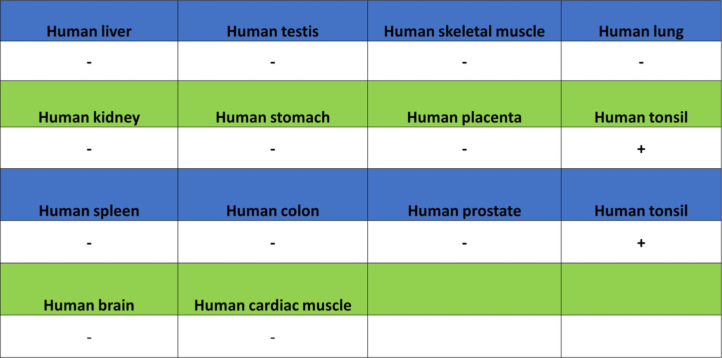
IHC shows positive staining in paraffin-embedded human tonsil. Anti-MEF2B antibody was used at 1/200 dilution, followed by a HRP Polymer for Rabbit IgG (ready to use). Counterstained with hematoxylin. Heat mediated antigen retrieval with Tris/EDTA buffer pH9.0 was performed before commencing with IHC staining protocol.
Immunocytochemistry
ICC shows positive staining in Ramos cells (top panel) and negative staining in HeLa cells (below panel). Anti- MEF2B antibody was used at 1/500 dilution (Green) and incubated overnight at 4°C. Goat polyclonal Antibody to Rabbit IgG - H&L (Alexa Fluor® 488) was used as secondary antibody at 1/1000 dilution. The cells were fixed with 4% PFA and permeabilized with 0.1% PBS-Triton X-100. Nuclei were counterstained with DAPI (Blue). Counterstain with tubulin (Red).