WB result of Mcl-1 Recombinant Rabbit mAb
Primary antibody: Mcl-1 Recombinant Rabbit mAb at 1/1000 dilution
Lane 1: L-929 whole cell lysate 20 µg
Lane 2: NIH/3T3 whole cell lysate 20 µg
Lane 3: C2C12 whole cell lysate 20 µg
Lane 4: mouse spleen lysate 20 µg
Secondary antibody: Goat Anti- rabbit IgG, (H+L), HRP conjugated at 1/10000 dilution
Predicted MW: 35 kDa
Observed MW: 36, 38 kDa
Mcl-1 Recombinant Rabbit mAb (S-3887-64)
Mcl-1 Recombinant Rabbit mAb (S-3887-64)
Price:
Regular price
$100 USD
Regular price
Sale price
$100 USD
Unit price
per
For shipping services or bulk orders, you may request a quotation.
Secure checkout with

View full details
Product Details
Product Details
Product Specification
| Host | Rabbit |
| Antigen | MCL1 |
| Synonyms | Induced myeloid leukemia cell differentiation protein Mcl-1 homolog; Bcl-2-related protein EAT/mcl1; Mcl1 |
| Immunogen | Synthetic Peptide |
| Location | Cytoplasm, Nucleus, Mitochondrion |
| Accession | P97287 |
| Clone Number | S-3887-64 |
| Antibody Type | Recombinant mAb |
| Isotype | IgG |
| Application | WB, ICC |
| Reactivity | Ms, Rt |
| Positive Sample | L929, NIH/3T3, C2C12, mouse spleen, C6, rat spleen |
| Purification | Protein A |
| Concentration | 0.5 mg/ml |
| Conjugation | Unconjugated |
| Physical Appearance | Liquid |
| Storage Buffer | PBS, 40% Glycerol, 0.05% BSA, 0.03% Proclin 300 |
| Stability & Storage | 12 months from date of receipt / reconstitution, -20 °C as supplied |
Dilution
| application | dilution | species |
| WB | 1:1000 | Ms, Rt |
| ICC | 1:500 | Ms |
Background
MCL1 (Myeloid cell leukemia 1) is an anti-apoptotic member of the BCL-2 protein family that plays a crucial role in regulating cell survival and death by binding to and neutralizing pro-apoptotic proteins such as BAK and BIM, thereby preventing mitochondrial outer membrane permeabilization and subsequent activation of the intrinsic apoptotic pathway.
Picture
Picture
Western Blot
WB result of Mcl-1 Recombinant Rabbit mAb
Primary antibody: Mcl-1 Recombinant Rabbit mAb at 1/1000 dilution
Lane 1: C6 whole cell lysate 20 µg
Lane 2: rat spleen lysate 20 µg
Secondary antibody: Goat Anti- rabbit IgG, (H+L), HRP conjugated at 1/10000 dilution
Predicted MW: 35 kDa
Observed MW: 36, 38 kDa
Immunocytochemistry
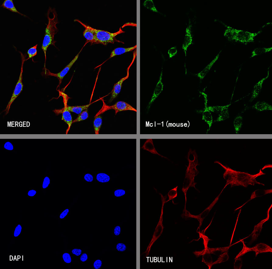
ICC shows positive staining in NIH/3T3 cells. Anti- Mcl-1 antibody was used at 1/500 dilution (Green) and incubated overnight at 4°C. Goat polyclonal Antibody to Rabbit IgG - H&L (Alexa Fluor® 488) was used as secondary antibody at 1/1000 dilution. The cells were fixed with 100% ice-cold methanol and permeabilized with 0.1% PBS-Triton X-100. Nuclei were counterstained with DAPI (Blue). Counterstain with tubulin (Red).