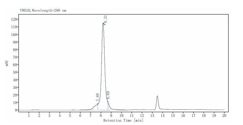
2μg (R: reducing condition).
MAT1A His Tag Protein, Human
MAT1A His Tag Protein, Human
Price:
Regular price
$270 USD
Regular price
Sale price
$270 USD
Unit price
per
For shipping services or bulk orders, you may request a quotation.
Secure checkout with

View full details
Product Details
Product Details
Product Specification
| Species | Human |
| Synonyms | AMS1,METK1 |
| Accession | Q00266 |
| Amino Acid Sequence | Met1-Phe395 with His&Thrombin Tag at the N-Terminus |
| Expression System | E. coli |
| Molecular Weight | 40-55kDa (Reducing) |
| Endotoxin | <1EU/μg |
| Tag | His Tag |
| Physical Appearance | Liquid |
| Storage Buffer | 20mM Tris-HCl pH-8, 1mM DTT, 100mM NaCl and 10% glycerol |
| Reconstitution | Reconstitute at 0.1-1 mg/ml according to the size in ultrapure water after rapid centrifugation |
| Stability & Storage | Stable for 12 months upon stored at -80℃ from the date of receipt. And avoid repeated freeze-thaws cycles. |
Background
MAT1A (methionine adenosyltransferase 1A) is mainly expressed in the liver, and the protein it encodes exists as dimers or tetramers, with each subunit containing approximately 395 amino acids and possessing a typical Rossmann-fold nucleotide-binding domain and a catalytic domain. Functionally, MAT1A catalyzes the synthesis of S-adenosylmethionine (SAM) from methionine and ATP, serving as a core enzyme in hepatic one-carbon metabolism and methylation reactions. Clinically, MAT1A is often silenced in hepatocellular carcinoma and cholangiocarcinoma due to promoter hypermethylation, leading to SAM depletion, DNA hypomethylation, and genomic instability that promote tumor progression; the expression ratio of MAT1A to MAT2A can serve as a prognostic marker for liver cancer, and restoring MAT1A expression or supplementing SAM holds therapeutic potential. In addition, MAT1A mutations can cause Mudd disease (hypermethioninemia), while alcohol promotes its degradation through phosphorylation and SUMOylation, contributing to the pathogenesis of alcoholic liver disease.
Protocol
Assay protocol
Principle:The MAT1A adenosyltransferase activity is detected in a colorimetric assay wherein the monophosphate generated from the enzyme reaction forms a colored complex with the Malachite Green Phosphate Detection Kit. The amount of complex can be then measured by a microplate reader.
Materials
1.MAT1A Dilution Buffer: 20mM Tris-HCl, pH8.0
2.MAT1A Assay Buffer (5X): 100mM Tris-HCl, pH8.0, 20mM MgCl2, 150mM KCl, 1mM DTT
3.MAT1A Assay Buffer (1X): 20mM Tris-HCl, pH8.0, 4mM MgCl2, 30mM KCl, 200uM DTT
4.MAT1A His Tag Protein, Human (Catalog # UA085073)
5.Malachite Green Phosphate Detection Kit (R&D, Catalog # DY996)
6.Substrate Assay Solution: ATP + L-methionine (Sigma, Catalog # M9625)
7.96-well half-area black plate with clear bottom (GENEVER, Catalog # GM04-96M)
8.Plate Reader (PerkinElmer)
Produce
1.Prepare a Phosphate Standard curve by adding 10 µL of the 1 M Phosphate Standard to 990 µL of MAT1A Assay Buffer (1X) for 10 mM stock. Continue by adding 10 µL of the 10 mM phosphate stock to 990 µL of MAT1A Assay Buffer (1X) for 100 µM stock. (This is the first dilution to use as a standard).
2.Perform six additional one-half dilutions of the 100 µM Phosphate stock.
3.Load 25 µL of each standard dilution to empty wells of a clear plate. Include a curve blank containing 25 µL MAT1A Assay Buffer (1X). The standard curve has a range of 0.039-2.5 nmol per well.
4.Thaw the Active MAT1A on ice. Prepare serial dilutions of MAT1A (40 µg/mL, 20 µg/mL, 10 µg/mL) using MAT1A Dilution Buffer.
5.Substrate Assay Solution: ATP solution, 10 mM; L-methionine, 150 mM.
Prepare ATP and L-methionine diluted in distilled water to a working concentration of 2.5 mM ATP / 5 mM L-methionine, resulting in a final concentration of 1 mM ATP / 2 mM L-methionine.
6.Add the following reaction components to bring the total volume to 25 µL:
Component 1: 10 µL of diluted Active MAT1A (Catalog # UA085073).
Component 2: 10 µL of Substrate Assay Solution.
Component 3. 5 µL 5X MAT1A Assay Buffer
7.Set up the blank control as outlined in step 6, excluding the addition of the Active Enzyme. Replace the MAT1A with an equal volume of MAT1A Dilution Buffer.
8.Cover the plate with a plate seal and incubate the mixture in a 37℃ incubator for 60 minutes.
9.After incubation, add 30 µL of Malachite Green Reagent A to all wells used. Mix briefly.
10.Add 100 μL of deionized water to all wells. Mix briefly.
11.Add 30 µL of Malachite Green Reagent B to all wells. Mix and incubate at room temperature for 20 minutes.
12.Read the plate at 620 nm in endpoint mode.
Specific Activity (nmol/min/mg) = |
Corresponding phosphate released (nmol) - Blank |
Incubation time (min) × amount of enzyme (mg) |
Experimental method
Experimental principleThe adenosyltransferase activity of MAT1A is detected by a colorimetric method. The monophosphate generated during the enzymatic reaction can form a colored complex with the Malachite Green Phosphate Detection Kit reagent, and the amount of this complex can be measured using a microplate reader.
Experimental materials
1.MAT1A Dilution Buffer: 20 mM Tris-HCl, pH 8.0
2.MAT1A Assay Buffer (5X): 100 mM Tris-HCl, pH 8.0, 20 mM MgCl₂2, 150 mM KCl, 1 mM DTT
3.MAT1A Assay Buffer (1X): 20 mM Tris-HCl, pH 8.0, 4 mM MgCl₂2, 30mM KCl, 200uM DTT
4.MAT1A His Tag Protein, Human (Catalog # UA085073)
5.Malachite Green Phosphate Detection Kit (R&D, Catalog # DY996)
6.Substrate: ATP + L-methionine (Sigma, Catalog # M9625)
7.96-well half-area black plate with clear bottom (GENEVER, Catalog # GM04-96M)
8.Plate reader (PerkinElmer)
Experimental procedure
1.Prepare the phosphate standard curve: Add 10 µL of 1 M phosphate standard solution to 990 µL of MAT1A assay buffer (1X) to prepare a 10 mM stock solution. Then, add 10 µL of the 10 mM phosphate stock solution to 990 µL of MAT1A assay buffer (1X) to prepare a 100 µM stock solution. (This serves as the first-level dilution used as the standard.)
2.Perform six serial half-fold dilutions of the 100 µM phosphate stock solution.
3.Take 25 µL of each concentration of the standard dilutions and add them to the wells of a clear microplate. Also set up a blank well containing 25 µL of MAT1A assay buffer (1X). The concentration range of the standard curve is 0.039–2.5 nmol per well.
4.Thaw the active MAT1A on ice. Prepare a series of dilutions of MAT1A (40 µg/mL, 20 µg/mL, 10 µg/mL) using the MAT1A dilution buffer.
5.Substrate detection solution: ATP solution, 10 mM; L-methionine, 150 mM.
Dilute ATP and L-methionine with distilled water to achieve a working concentration of 2.5 mM ATP / 5 mM L-methionine, resulting in final concentrations of 1 mM ATP / 2 mM L-methionine.
6.Add the following reaction components to bring the total volume to 25 µl:
Component 1: 10 µl of diluted active MAT1A (Cat. No. UA085073)
Component 2: 10 µl of substrate detection solution
Component 3. 5 µl of 5X MAT1A assay buffer
7.Set up the blank control as described in step 6, but do not add MAT1A. Replace MAT1A with an equal volume of MAT1A dilution buffer.
8.Cover the microplate with a plate lid and incubate the mixture in a 37°C incubator for 60 minutes.
9.After incubation, add 30 µL of Malachite Green Reagent A to all wells used, and mix gently.
10.Add 100 µL of deionized water to all wells, and mix gently.
11.Add 30 µL of Malachite Green Reagent B to all wells. After mixing, incubate at room temperature for 20 minutes.
12.Read the microplate in endpoint mode at a wavelength of 620 nm.
Specific Activity (nmol/min/mg) = |
Corresponding phosphate released (nmol) - Blank |
Incubation time (min) × amount of enzyme (mg) |
Picture
Picture
SDS-PAGE
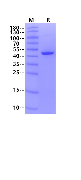
SEC-HPLC
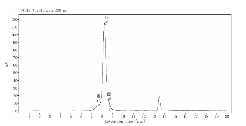
The purity of MAT1A His Tag Protein, Human is more than 85% as determined by SEC-HPLC.