WB result of LPCAT2 Recombinant Rabbit mAb
Primary antibody: LPCAT2 Recombinant Rabbit mAb at 1/1000 dilution
Lane 1: Raji whole cell lysate 20 µg
Lane 2: SW480 whole cell lysate 20 µg
Negative control: Raji whole cell lysate
Secondary antibody: Goat Anti-Rabbit IgG (H+L), HRP conjugated at 1/10000 dilution
Predicted MW: 60 kDa
Observed MW: 50 kDa
LPCAT2 Recombinant Rabbit mAb (S-4239-18)
LPCAT2 Recombinant Rabbit mAb (S-4239-18)
Price:
Regular price
$100 USD
Regular price
Sale price
$100 USD
Unit price
per
For shipping services or bulk orders, you may request a quotation.
Secure checkout with

View full details
Product Details
Product Details
Product Specification
| Host | Rabbit |
| Antigen | LPCAT2 |
| Synonyms | Lysophosphatidylcholine acyltransferase 2; LPC acyltransferase 2; LPCAT-2; LysoPC acyltransferase 2; 1-acylglycerol-3-phosphate O-acyltransferase 11 (1-AGP acyltransferase 11; 1-AGPAT 11); 1-acylglycerophosphocholine O-acyltransferase; 1-alkenylglycerophosphocholine O-acyltransferase; 1-alkylglycerophosphocholine O-acetyltransferase; Acetyl-CoA:lyso-platelet-activating factor acetyltransferase (Acetyl-CoA:lyso-PAF acetyltransferase; Lyso-PAF acetyltransferase; LysoPAFAT); AGPAT11; AYTL1 |
| Immunogen | Synthetic Peptide |
| Location | Cell membrane, Endoplasmic reticulum |
| Accession | Q7L5N7 |
| Clone Number | S-4239-18 |
| Antibody Type | Recombinant mAb |
| Isotype | IgG |
| Application | WB, IHC-P, ICC |
| Reactivity | Hu, Ms, Rt, Mk |
| Positive Sample | SW480, Jurkat, A431, mouse liver, PC-12, COS7 |
| Purification | Protein A |
| Concentration | 0.5 mg/ml |
| Conjugation | Unconjugated |
| Physical Appearance | Liquid |
| Storage Buffer | PBS, 40% Glycerol, 0.05% BSA, 0.02% sodium azide |
| Stability & Storage | 12 months from date of receipt / reconstitution, -20 °C as supplied |
Dilution
| application | dilution | species |
| WB | 1:1000 | Hu, Ms, Rt, Mk |
| IHC-P | 1:200 | Hu, Ms, Rt |
| ICC | 1:500 | Hu |
Background
Lysophosphatidylcholine acyltransferase 2 (LPCAT2) is a membrane-bound enzyme belonging to the lysophospholipid acyltransferase family that catalyzes the reacylation of lysophosphatidylcholine to phosphatidylcholine using acyl-CoA donors, thereby playing a pivotal role in the Lands cycle of phospholipid remodeling and maintaining cellular membrane homeostasis. Beyond its fundamental metabolic function, LPCAT2 is critically implicated in inflammatory responses and cancer biology, as it is frequently upregulated in various malignancies including colorectal, breast, and hepatocellular carcinomas, where its overexpression promotes tumor cell proliferation, migration, and invasion through the activation of signaling pathways such as PI3K/AKT and MAPK/ERK while also modulating the composition of lipid rafts to influence receptor tyrosine kinase activity. Furthermore, LPCAT2 has been identified as a key mediator in macrophage activation and foam cell formation during atherosclerosis, linking lipid metabolism directly to innate immunity, which collectively positions this enzyme as both a potential prognostic biomarker and a therapeutic target for treating LPCAT2-dependent cancers and chronic inflammatory diseases.
Picture
Picture
Western Blot
WB result of LPCAT2 Recombinant Rabbit mAb
Primary antibody: LPCAT2 Recombinant Rabbit mAb at 1/1000 dilution
Lane 1: Jurkat whole cell lysate 20 µg
Lane 2: A431 whole cell lysate 20 µg
Secondary antibody: Goat Anti-Rabbit IgG (H+L), HRP conjugated at 1/10000 dilution
Predicted MW: 60 kDa
Observed MW: 50 kDa
WB result of LPCAT2 Recombinant Rabbit mAb
Primary antibody: LPCAT2 Recombinant Rabbit mAb at 1/1000 dilution
Lane 1: mouse liver lysate 20 µg
Secondary antibody: Goat Anti-Rabbit IgG (H+L), HRP conjugated at 1/10000 dilution
Predicted MW: 60 kDa
Observed MW: 50 kDa
WB result of LPCAT2 Recombinant Rabbit mAb
Primary antibody: LPCAT2 Recombinant Rabbit mAb at 1/1000 dilution
Lane 1: PC-12 whole cell lysate 20 µg
Secondary antibody: Goat Anti-Rabbit IgG (H+L), HRP conjugated at 1/10000 dilution
Predicted MW: 60 kDa
Observed MW: 50 kDa
WB result of LPCAT2 Recombinant Rabbit mAb
Primary antibody: LPCAT2 Recombinant Rabbit mAb at 1/1000 dilution
Lane 1: COS7 whole cell lysate 20 µg
Secondary antibody: Goat Anti-Rabbit IgG (H+L), HRP conjugated at 1/10000 dilution
Predicted MW: 60 kDa
Observed MW: 50 kDa
Immunohistochemistry
IHC shows positive staining in paraffin-embedded human cerebral cortex. Anti-LPCAT2 antibody was used at 1/200 dilution, followed by a HRP Polymer for Rabbit IgG (ready to use). Counterstained with hematoxylin. Heat mediated antigen retrieval with Tris/EDTA buffer pH9.0 was performed before commencing with IHC staining protocol.
IHC shows positive staining in paraffin-embedded human colon. Anti-LPCAT2 antibody was used at 1/200 dilution, followed by a HRP Polymer for Rabbit IgG (ready to use). Counterstained with hematoxylin. Heat mediated antigen retrieval with Tris/EDTA buffer pH9.0 was performed before commencing with IHC staining protocol.
IHC shows positive staining in paraffin-embedded human spleen. Anti-LPCAT2 antibody was used at 1/200 dilution, followed by a HRP Polymer for Rabbit IgG (ready to use). Counterstained with hematoxylin. Heat mediated antigen retrieval with Tris/EDTA buffer pH9.0 was performed before commencing with IHC staining protocol.
IHC shows positive staining in paraffin-embedded human testis. Anti-LPCAT2 antibody was used at 1/200 dilution, followed by a HRP Polymer for Rabbit IgG (ready to use). Counterstained with hematoxylin. Heat mediated antigen retrieval with Tris/EDTA buffer pH9.0 was performed before commencing with IHC staining protocol.
Negative control: IHC shows negative staining in paraffin-embedded human skeletal muscle. Anti-LPCAT2 antibody was used at 1/200 dilution, followed by a HRP Polymer for Rabbit IgG (ready to use). Counterstained with hematoxylin. Heat mediated antigen retrieval with Tris/EDTA buffer pH9.0 was performed before commencing with IHC staining protocol.
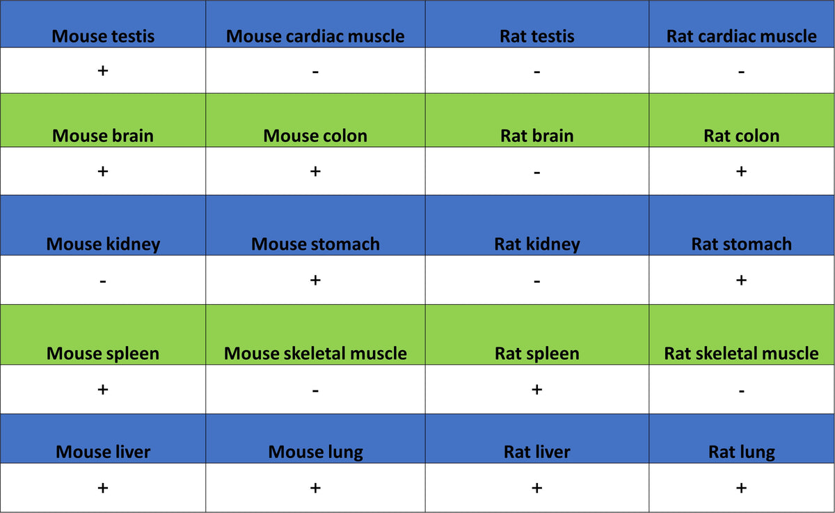
IHC shows positive staining in paraffin-embedded mouse spleen. Anti-LPCAT2 antibody was used at 1/200 dilution, followed by a HRP Polymer for Rabbit IgG (ready to use). Counterstained with hematoxylin. Heat mediated antigen retrieval with Tris/EDTA buffer pH9.0 was performed before commencing with IHC staining protocol.
IHC shows positive staining in paraffin-embedded rat spleen. Anti-LPCAT2 antibody was used at 1/200 dilution, followed by a HRP Polymer for Rabbit IgG (ready to use). Counterstained with hematoxylin. Heat mediated antigen retrieval with Tris/EDTA buffer pH9.0 was performed before commencing with IHC staining protocol.
Immunocytochemistry
ICC shows positive staining in SW480 cells (top panel) and negative staining in Raji cells (below panel). Anti-LPCAT2 antibody was used at 1/500 dilution (Green) and incubated overnight at 4°C. Goat polyclonal Antibody to Rabbit IgG - H&L (Alexa Fluor® 488) was used as secondary antibody at 1/1000 dilution. The cells were fixed with 4% PFA and permeabilized with 0.1% PBS-Triton X-100. Nuclei were counterstained with DAPI (Blue). Counterstain with tubulin (Red).