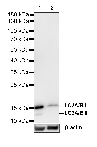
LC3A/B-Knockout HeLa Cell Lysate
WB result:
Primary antibody: LC3A/B Recombinant Rabbit mAb (S0B1237) at 1/1000 dilution
Lane 1: Wild-type HeLa whole cell lysate 20 μg
Lane 2: LC3A/B-Knockout HeLa whole Cell lysate 20 μg
Secondary antibody: Goat Anti-Rabbit lgG (H+L), HRP conjugated at 1/10000 dilution
Predicted band size: 14, 15 kDa
Observed band size: 14, 15 kDa
LC3A/B-Knockdown HeLa Cell Lysate
LC3A/B-Knockdown HeLa Cell Lysate
Price:
Regular price
$670 USD
Regular price
Sale price
$670 USD
Unit price
per
For shipping services or bulk orders, you may request a quotation.
Secure checkout with

View full details
Product Details
Product Details
Product Specification
| Host | Human |
| Application | WB |
| Concentration | 2 mg/ml |
| Physical Appearance | Liquid |
| Storage Buffer | Supplied in SDS Sample Buffer: 50 mM Tris-HCl (pH 6.8 at 25°C), 2% w/v SDS, 10% glycerol, 1% v/v β-Mercaptoethanol, 0.01% w/v bromophenol blue or phenol red. |
| Stability & Storage | -20 °C for 1 months under sterile conditions; |
Background
Microtubule-associated protein 1 light chain 3 (LC3), encoded by MAP1LC3A (LC3A) and MAP1LC3B (LC3B), represents a mammalian homolog of yeast Atg8 and serves as a fundamental structural constituent and canonical marker of macroautophagy. Synthesized as precursor proteins, LC3 isoforms are initially cleaved at their C-termini by the cysteine protease ATG4B to expose a conserved glycine residue, yielding the cytosolic form designated LC3-I. Upon induction of autophagy by nutrient starvation, proteotoxic stress, organelle damage, or pathogen invasion, LC3-I is covalently conjugated to phosphatidylethanolamine (PE) via an ubiquitin-like cascade involving ATG7 (E1-like) and ATG3 (E2-like) enzymes together with the ATG12–ATG5–ATG16L1 complex (E3-like), generating the lipidated form termed LC3-II. Lipidated LC3-II specifically integrates into both the inner and outer membranes of expanding phagophores and mature autophagosomes, orchestrating membrane elongation, closure, and cargo sequestration through direct interaction with selective autophagy receptors (such as p62/SQSTM1 and NBR1) via conserved LC3-interacting regions (LIRs). Following autophagosome-lysosome fusion, intra-autophagosomal LC3-II is degraded by lysosomal hydrolases, rendering the dynamic conversion of LC3-I to LC3-II and the subsequent turnover of LC3-II reliable surrogate indicators of autophagic flux. LC3A/B antibodies—frequently designed to co-detect both isoforms or specifically discriminate LC3B—are widely utilized in Western blotting (evaluating the LC3-II/LC3-I ratio in the presence and absence of lysosomal inhibitors such as bafilomycin A1 or chloroquine), immunofluorescence, and immunohistochemistry (monitoring puncta formation corresponding to autophagic vesicles). Knockout (KO) of LC3A and/or LC3B (MAP1LC3A/MAP1LC3B) can impair autophagosome biogenesis, disrupt selective cargo recruitment, and attenuate autophagic degradation under stress conditions, potentially sensitizing cells to proteotoxicity and metabolic starvation; however, the overt cellular defect can be partially mitigated by functional redundancy and compensatory actions of other mammalian Atg8 family homologs, notably the GABARAP subfamily (GABARAP, GABARAPL1, and GABARAPL2).
Picture
Picture
Western Blot