WB result of KLC1 Recombinant Rabbit mAb
Primary antibody: KLC1 Recombinant Rabbit mAb at 1/1000 dilution
Lane 1: 293T whole cell lysate 20 µg
Lane 2: HeLa whole cell lysate 20 µg
Lane 3: U-87 MG whole cell lysate 20 µg
Secondary antibody: Goat Anti-Rabbit IgG (H+L), HRP conjugated at 1/10000 dilution
Predicted MW: 65 kDa
Observed MW: 65, 75 kDa
KLC1 Recombinant Rabbit mAb (S-4556-8)
KLC1 Recombinant Rabbit mAb (S-4556-8)
Price:
Regular price
$100 USD
Regular price
Sale price
$100 USD
Unit price
per
For shipping services or bulk orders, you may request a quotation.
Secure checkout with

View full details
Product Details
Product Details
Product Specification
| Host | Rabbit |
| Antigen | KLC1 |
| Synonyms | Kinesin light chain 1; KLC; KNS2 |
| Immunogen | Synthetic Peptide |
| Location | Cytoplasm, Cytoskeleton |
| Accession | Q07866 |
| Clone Number | S-4556-8 |
| Antibody Type | Recombinant mAb |
| Isotype | IgG |
| Application | WB, IHC-P, ICC |
| Reactivity | Hu, Ms, Rt |
| Positive Sample | 293T, HeLa, U-87 MG, Neuro-2a, C2C12, mouse brain, PC-12, rat brain, COS-7 |
| Predicted Reactivity | Or |
| Purification | Protein A |
| Concentration | 0.5 mg/ml |
| Conjugation | Unconjugated |
| Physical Appearance | Liquid |
| Storage Buffer | PBS, 40% Glycerol, 0.05% BSA, 0.02% sodium azide |
| Stability & Storage | 12 months from date of receipt / reconstitution, -20 °C as supplied |
Dilution
| application | dilution | species |
| WB | 1:1000 | Hu, Ms, Rt, Mk |
| IHC-P | 1:1000 | Hu, Ms, Rt |
| ICC | 1:500 | Hu, Ms |
Background
KLC1 (kinesin light chain 1) is the light chain subunit of conventional kinesin-1, encoded by the KLC1 gene, and is predominantly expressed in neuronal tissues. Kinesin-1 is a heterotetramer composed of two heavy chains (KIF5A/B/C) and two light chains, in which the heavy chains generate the force for movement along microtubules toward the plus end, while the light chains themselves are not motor proteins but primarily serve as adaptor molecules. They bind to the heavy chains through their N-terminal domain and interact with a variety of cargoes—including vesicles, mitochondria, and the Golgi apparatus—via six tetratricopeptide repeat (TPR) motifs. The function of KLC1 is particularly critical for neurons: KLC1 knockout mice are significantly smaller in size and exhibit pronounced motor impairments, with KIF5A abnormally co-localizing with cis-Golgi markers in their sensory and motor neurons, and the intracellular distribution of KIF5B and β'-COP also being markedly altered. These findings indicate that KLC1 is essential for the proper activation or targeting of kinesin heavy chains. Furthermore, KLC1 also interacts with specific cargo receptors such as the mitochondrial outer membrane protein FUNDC1 through its TPR domain, further expanding its adaptor functions in intracellular material transport.
Picture
Picture
Western Blot
WB result of KLC1 Recombinant Rabbit mAb
Primary antibody: KLC1 Recombinant Rabbit mAb at 1/1000 dilution
Lane 1: Neuro-2a whole cell lysate 20 µg
Lane 2: C2C12 whole cell lysate 20 µg
Lane 3: mouse brain lysate 20 µg
Secondary antibody: Goat Anti-Rabbit IgG (H+L), HRP conjugated at 1/10000 dilution
Predicted MW: 65 kDa
Observed MW: 65, 75 kDa
WB result of KLC1 Recombinant Rabbit mAb
Primary antibody: KLC1 Recombinant Rabbit mAb at 1/1000 dilution
Lane 1: PC-12 whole cell lysate 20 µg
Lane 2: rat brain lysate 20 µg
Secondary antibody: Goat Anti-Rabbit IgG (H+L), HRP conjugated at 1/10000 dilution
Predicted MW: 65 kDa
Observed MW: 65, 75 kDa
WB result of KLC1 Recombinant Rabbit mAb
Primary antibody: KLC1 Recombinant Rabbit mAb at 1/1000 dilution
Lane 1: COS-7 whole cell lysate 20 µg
Secondary antibody: Goat Anti-Rabbit IgG (H+L), HRP conjugated at 1/10000 dilution
Predicted MW: 65 kDa
Observed MW: 65, 75 kDa
Immunohistochemistry
IHC shows positive staining in paraffin-embedded human brain. Anti-KLC1 antibody was used at 1/1000 dilution, followed by a HRP Polymer for Rabbit IgG (ready to use). Counterstained with hematoxylin. Heat mediated antigen retrieval with Tris/EDTA buffer pH9.0 was performed before commencing with IHC staining protocol.
IHC shows positive staining in paraffin-embedded human breast cancer. Anti-KLC1 antibody was used at 1/1000 dilution, followed by a HRP Polymer for Rabbit IgG (ready to use). Counterstained with hematoxylin. Heat mediated antigen retrieval with Tris/EDTA buffer pH9.0 was performed before commencing with IHC staining protocol.
IHC shows positive staining in paraffin-embedded mouse brain. Anti-KLC1 antibody was used at 1/1000 dilution, followed by a HRP Polymer for Rabbit IgG (ready to use). Counterstained with hematoxylin. Heat mediated antigen retrieval with Tris/EDTA buffer pH9.0 was performed before commencing with IHC staining protocol.
IHC shows positive staining in paraffin-embedded rat brain. Anti-KLC1 antibody was used at 1/1000 dilution, followed by a HRP Polymer for Rabbit IgG (ready to use). Counterstained with hematoxylin. Heat mediated antigen retrieval with Tris/EDTA buffer pH9.0 was performed before commencing with IHC staining protocol.
Immunocytochemistry
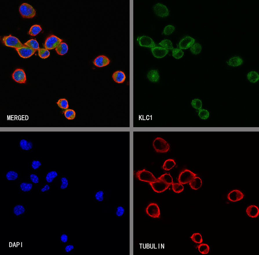
ICC shows positive staining in HeLa cells. Anti- KLC1 antibody was used at 1/500 dilution (Green) and incubated overnight at 4°C. Goat polyclonal Antibody to Rabbit IgG - H&L (Alexa Fluor® 488) was used as secondary antibody at 1/1000 dilution. The cells were fixed with 4% PFA and permeabilized with 0.1% PBS-Triton X-100. Nuclei were counterstained with DAPI (Blue). Counterstain with tubulin (Red).
ICC shows positive staining in Neuro-2a cells. Anti- KLC1 antibody was used at 1/500 dilution (Green) and incubated overnight at 4°C. Goat polyclonal Antibody to Rabbit IgG - H&L (Alexa Fluor® 488) was used as secondary antibody at 1/1000 dilution. The cells were fixed with 4% PFA and permeabilized with 0.1% PBS-Triton X-100. Nuclei were counterstained with DAPI (Blue). Counterstain with tubulin (Red).