Skip to product information
1 of 10

JUND Recombinant Rabbit mAb (S-4270-48)

JUND Recombinant Rabbit mAb (S-4270-48)

Catalog Number: S0B60689 Application: WB, IHC-P, ICC Reactivity: Hu, Ms, Rt, Mk Conjugation: Unconjugated Brand: Starter
Price:
Regular price $100 USD
Regular price Sale price $100 USD
Size:

For shipping services or bulk orders, you may request a quotation.
Secure checkout with
View full details

Product Details

Product Specification


Host Rabbit
Synonyms Transcription factor JunD; Transcription factor AP-1 subunit JunD
Immunogen Synthetic Peptide
Location Nucleus
Accession P17535
Clone Number S-4270-48
Antibody Type Recombinant mAb
Isotype IgG
Application WB, IHC-P, ICC
Reactivity Hu, Ms, Rt, Mk
Predicted Reactivity Bv
Purification Protein A
Concentration 0.25 mg/ml
Expression System Rabbit
Conjugation Unconjugated
Physical Appearance Liquid
Storage Buffer

PBS, 40% Glycerol, 0.05% BSA, 0.02% sodium azide

Stability & Storage

12 months from date of receipt / reconstitution,-20 °C as supplied

Dilution


application dilution species
WB 1:500-1:1000 Hu, Ms, Rt, Mk
IHC-P 1:500-1:2500 Hu, Ms, Rt
ICC 1:250 Hu, Ms

Background

JUND (JunD transcription factor) is a basic region leucine zipper (bZIP)transcription factor encoded by the JUND gene located on human chromosome19p13.11, and is a core component of the AP-1 transcription factor complex. Theprotein contains 347 amino acids with a molecular weight of approximately 35.2kDa, and from the N-terminus to the C-terminus it sequentially comprises a JNKphosphorylation motif, a DNA-binding domain, a nuclear localization signal, anda leucine zipper domain. Due to the lack of a JNK docking site, JUND can onlybe weakly phosphorylated by JNK. JUND is the most widely expressed butlowest-abundance member of the JUN family; although its mRNA is a singletranscript, alternative in-frame translation initiation sites produce twoisoforms, JUND-L and JUND-S, of which JUND-S lacks 43 N-terminal amino acidsand is therefore unable to bind the tumor suppressor protein Menin.Functionally, JUND forms heterodimers with FOS family proteins and binds theAP-1 consensus sequence to regulate transcription. In contrast to JUN, whoseexpression decreases when cells enter G0 phase, JUND expression increases underserum starvation-induced quiescence and acts as an antagonist of JUN byinhibiting cyclin D1 induction, thereby helping to maintain the quiescent stateof cells. In addition, JUND exerts anti-angiogenic effects by regulatingRas-mediated reactive oxygen species production and protects cells fromp53-dependent senescence and apoptosis. JUND-deficient mice are viable andexhibit only mild defects in growth and spermatogenesis, in striking contrastto the embryonic lethality caused by JUN or JUNB deletion. In terms of diseaseassociations, JUND has been linked to T cell leukemia and multiple endocrineneoplasia type 1, and plays important roles in macrophage activation andsusceptibility to glomerulonephritis.

Picture

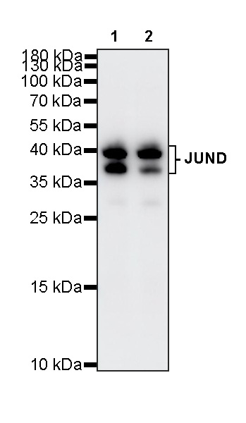
Western Blot

WB result of JUND Recombinant Rabbit mAb

Primary antibody: JUND Recombinant Rabbit mAb at 1/1000 dilution

Lane 1: HeLa whole cell lysate 20 μg

Lane 2: K-562 whole cell lysate 20 μg

Secondary antibody: Goat Anti-Rabbit IgG (H+L), HRP conjugated at 1/10000 dilution

Predicted MW: 32 kDa

Observed MW: 37, 40 kDa
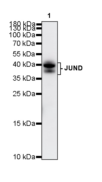
WB result of JUND Recombinant Rabbit mAb

Primary antibody: JUND Recombinant Rabbit mAb at 1/1000 dilution

Lane 1: NIH/3T3 whole cell lysate 20 μg

Secondary antibody: Goat Anti-Rabbit IgG (H+L), HRP conjugated at 1/10000 dilution

Predicted MW: 32 kDa

Observed MW: 37, 40 kDa
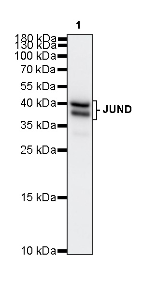
WB result of JUND Recombinant Rabbit mAb

Primary antibody: JUND Recombinant Rabbit mAb at 1/1000 dilution

Lane 1: C6 whole cell lysate 20 μg

Secondary antibody: Goat Anti-Rabbit IgG (H+L), HRP conjugated at 1/10000 dilution

Predicted MW: 32 kDa

Observed MW: 37, 40 kDa
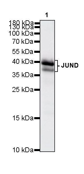
This blot was developed with high sensitivity substrate

WB result of JUND Recombinant Rabbit mAb

Primary antibody: JUND Recombinant Rabbit mAb at 1/1000 dilution

Lane 1: COS-7 whole cell lysate 20 μg

Secondary antibody: Goat Anti-Rabbit IgG (H+L), HRP conjugated at 1/10000 dilution

Predicted MW: 32 kDa

Observed MW: 37, 40 kDa
Immunohistochemistry

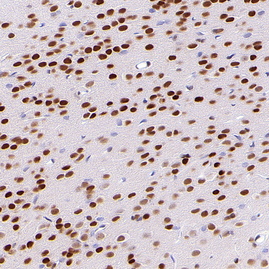
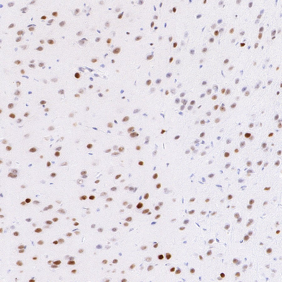
IHC shows positive staining in paraffin-embedded mouse brain. Anti-JUND antibody was used at 1/500 dilution, followed by a HRP Polymer for Mouse & Rabbit IgG (ready to use). Counterstained with hematoxylin. Heat mediated antigen retrieval with Tris/EDTA buffer pH9.0 was performed before commencing with IHC staining protocol.

IHC shows positive staining in paraffin-embedded rat brain. Anti-JUND antibody was used at 1/500 dilution, followed by a HRP Polymer for Mouse & Rabbit IgG (ready to use). Counterstained with hematoxylin. Heat mediated antigen retrieval with Tris/EDTA buffer pH9.0 was performed before commencing with IHC staining protocol.

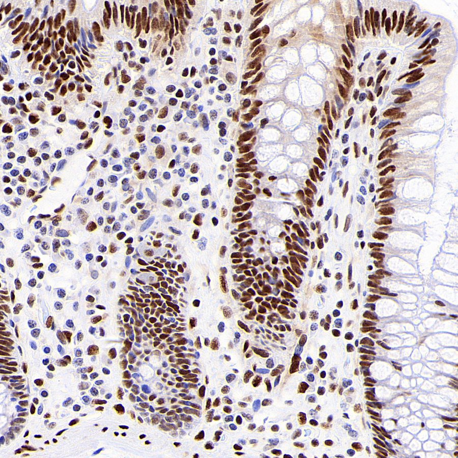
IHC shows positive staining in paraffin-embedded human colon. Anti-JUND antibody was used at 1/2500 dilution, followed by a HRP Polymer for Mouse & Rabbit IgG (ready to use). Counterstained with hematoxylin. Heat mediated antigen retrieval with Tris/EDTA buffer pH9.0 was performed before commencing with IHC staining protocol.

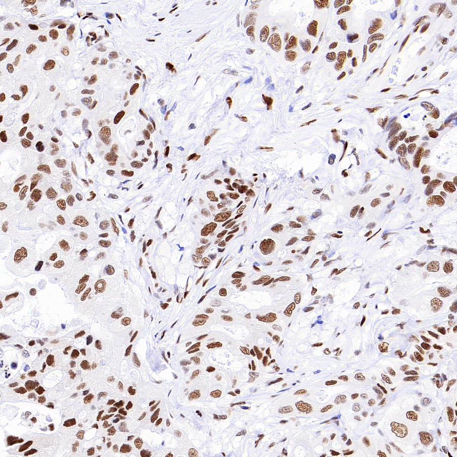
IHC shows positive staining in paraffin-embedded human gastric cancer. Anti-JUND antibody was used at 1/2500 dilution, followed by a HRP Polymer for Mouse & Rabbit IgG (ready to use). Counterstained with hematoxylin. Heat mediated antigen retrieval with Tris/EDTA buffer pH9.0 was performed before commencing with IHC staining protocol.

Immunocytochemistry

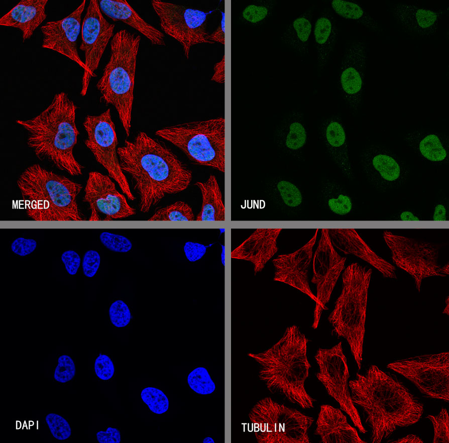
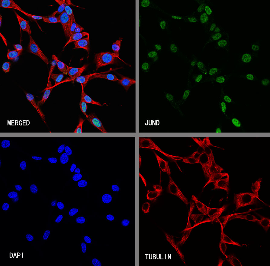
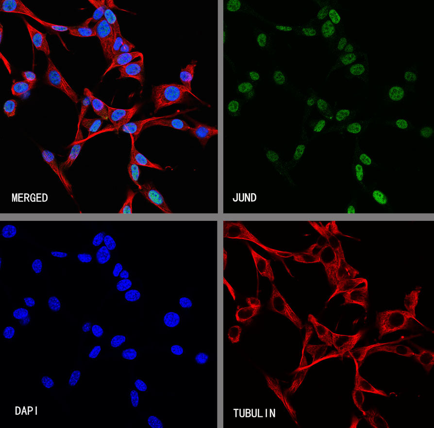
ICC shows positive staining in NIH/3T3 cells. Anti-JUND antibody was used at 1/250 dilution (Green) and incubated overnight at 4°C. Goat polyclonal Antibody to Rabbit IgG - H&L (Alexa Fluor® 488) was used as secondary antibody at 1/1000 dilution. The cells were fixed with 4% PFA and permeabilized with 0.1% PBS-Triton X-100. Nuclei were counterstained with DAPI (Blue). Counterstain with tubulin (Red).