WB result of IRP2 Recombinant Rabbit mAb
Primary antibody: IRP2 Recombinant Rabbit mAb at 1/1000 dilution
Lane 1: untreated HEK-293 whole cell lysate 20 μg
Lane 2: HEK-293 treated with 50μM fac overnight whole cell lysate 20 μg
Secondary antibody: Goat Anti-Rabbit IgG (H+L), HRP conjugated at 1/10000 dilution
Predicted MW: 105 kDa
Observed MW: 105 kDa
IRP2 Recombinant Rabbit mAb (S-4592-56)
IRP2 Recombinant Rabbit mAb (S-4592-56)
Price:
Regular price
$100 USD
Regular price
Sale price
$100 USD
Unit price
per
For shipping services or bulk orders, you may request a quotation.
Secure checkout with

View full details
Product Details
Product Details
Product Specification
| Host | Rabbit |
| Antigen | IRP2 |
| Synonyms | Iron-responsive element-binding protein 2; IREB2 |
| Immunogen | Synthetic Peptide |
| Location | Cytoplasm |
| Accession | P48200 |
| Clone Number | S-4592-56 |
| Antibody Type | Recombinant mAb |
| Isotype | IgG |
| Application | WB, ICC, IF |
| Reactivity | Hu, Ms, Rt |
| Positive Sample | A549, Jurkat, mouse kidney, mouse lung, rat kidney, rat lung |
| Purification | Protein A |
| Concentration | 0.5 mg/ml |
| Conjugation | Unconjugated |
| Physical Appearance | Liquid |
| Storage Buffer | PBS, 40% Glycerol, 0.05% BSA, 0.02% sodium azide |
| Stability & Storage | 12 months from date of receipt / reconstitution, -20 °C as supplied |
Dilution
| application | dilution | species |
| WB | 1:1000 | Hu, Ms, Rt |
| ICC | 1:500 | Hu |
| IF | 1:500 | Ms, Rt |
Background
IRP2 (Iron Regulatory Protein 2), encoded by the IREB2 gene, is a core regulator of intracellular iron homeostasis, functioning by binding to iron-responsive elements (IREs) on mRNAs to modulate the expression of iron metabolism-related genes. The activity of IRP2 is finely regulated by intracellular iron levels: when cellular iron is deficient, IRP2 remains stable through conformational changes in its iron-sulfur cluster-binding domain and binds with high affinity to IREs on target mRNAs, thereby suppressing the translation of ferritin and ferroportin while enhancing the stability of transferrin receptor 1 (TfR1) mRNA, collectively promoting iron uptake and reducing iron storage and export; conversely, when iron is abundant, IRP2 is degraded via the ubiquitin-proteasome pathway mediated by the FBXL5 E3 ubiquitin ligase, relieving translational repression of genes such as ferritin to safely store excess iron ions. Dysregulation of IRP2 disrupts systemic and cellular iron balance: its deficiency or loss of function in animal models leads to tissue iron overload and neurodegenerative disease-like phenotypes (such as axonal damage and motor deficits), and mutations in this gene have also been associated with human neurodegenerative diseases. Additionally, IRP2 plays a complex role in tumor progression by modulating iron metabolism in cancer cells (e.g., affecting iron uptake and reactive oxygen species levels), positioning it as a hotspot target in the research fields of ferroptosis, cancer, and neurodegenerative diseases.
Picture
Picture
Western Blot
WB result of IRP2 Recombinant Rabbit mAb
Primary antibody: IRP2 Recombinant Rabbit mAb at 1/1000 dilution
Lane 1: A549 whole cell lysate 20 μg
Lane 2: Jurkat whole cell lysate 20 μg
Secondary antibody: Goat Anti-Rabbit IgG (H+L), HRP conjugated at 1/10000 dilution
Predicted MW: 105 kDa
Observed MW: 105 kDa
WB result of IRP2 Recombinant Rabbit mAb
Primary antibody: IRP2 Recombinant Rabbit mAb at 1/1000 dilution
Lane 1: mouse kidney lysate 20 μg
Lane 2: mouse lung lysate 20 μg
Secondary antibody: Goat Anti-Rabbit IgG (H+L), HRP conjugated at 1/10000 dilution
Predicted MW: 105 kDa
Observed MW: 105 kDa
WB result of IRP2 Recombinant Rabbit mAb
Primary antibody: IRP2 Recombinant Rabbit mAb at 1/1000 dilution
Lane 1: rat kidney lysate 20 μg
Lane 2: rat lung lysate 20 μg
Secondary antibody: Goat Anti-Rabbit IgG (H+L), HRP conjugated at 1/10000 dilution
Predicted MW: 105 kDa
Observed MW: 105 kDa
Immunocytochemistry
ICC shows positive staining in A549 cells. Anti- IRP2 antibody was used at 1/500 dilution (Green) and incubated overnight at 4°C. Goat polyclonal Antibody to Rabbit IgG - H&L (Alexa Fluor® 488) was used as secondary antibody at 1/1000 dilution. The cells were fixed with 100% ice-cold methanol and permeabilized with 0.1% PBS-Triton X-100. Nuclei were counterstained with DAPI (Blue). Counterstain with tubulin (Red).
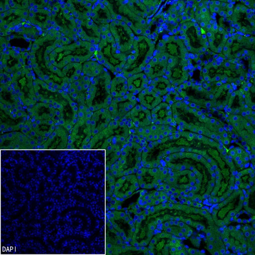
Immunofluorescence
IF shows positive staining in paraffin-embedded mouse kidney. Anti-IRP2 antibody was used at 1/500 dilution (Green) and incubated overnight at 4°C. Goat polyclonal Antibody to Rabbit IgG - H&L (Alexa Fluor® 488) was used as secondary antibody at 1/1000 dilution. Counterstained with DAPI (Blue). Heat mediated antigen retrieval with EDTA buffer pH9.0 was performed before commencing with IF staining protocol.
IF shows positive staining in paraffin-embedded rat kidney. Anti-IRP2 antibody was used at 1/500 dilution (Green) and incubated overnight at 4°C. Goat polyclonal Antibody to Rabbit IgG - H&L (Alexa Fluor® 488) was used as secondary antibody at 1/1000 dilution. Counterstained with DAPI (Blue). Heat mediated antigen retrieval with EDTA buffer pH9.0 was performed before commencing with IF staining protocol.