WB result of IRE1α Recombinant Rabbit mAb
Primary antibody: IRE1α Recombinant Rabbit mAb at 1/1000 dilution
Lane 1: Raji whole cell lysate 20 µg
Lane 2: HEK-293 whole cell lysate 20 µg
Lane 3: A549 whole cell lysate 20 µg
Lane 4: LNCaP whole cell lysate 20 µg
Lane 5: HeLa whole cell lysate 20 µg
Lane 6: Caco-2 whole cell lysate 20 µg
Secondary antibody: Goat Anti- rabbit IgG, (H+L), HRP conjugated at 1/10000 dilution
Predicted MW: 110 kDa
Observed MW: 130 kDa
IRE1α Recombinant Rabbit mAb (S-3873-25)
IRE1α Recombinant Rabbit mAb (S-3873-25)
Price:
Regular price
$100 USD
Regular price
Sale price
$100 USD
Unit price
per
For shipping services or bulk orders, you may request a quotation.
Secure checkout with

View full details
Product Details
Product Details
Product Specification
| Host | Rabbit |
| Antigen | IRE1α |
| Synonyms | Serine/threonine-protein kinase/endoribonuclease IRE1; Endoplasmic reticulum-to-nucleus signaling 1; Inositol-requiring protein 1 (hIRE1p); Ire1-alpha (IRE1a); IRE1; ERN1 |
| Immunogen | Synthetic Peptide |
| Location | Endoplasmic reticulum |
| Accession | O75460 |
| Clone Number | S-3873-25 |
| Antibody Type | Recombinant mAb |
| Isotype | IgG |
| Application | WB |
| Reactivity | Hu, Mk |
| Positive Sample | Raji, HEK-293, A549, LNCap, HeLa, Caco-2, COS-7 |
| Purification | Protein A |
| Concentration | 0.5 mg/ml |
| Conjugation | Unconjugated |
| Physical Appearance | Liquid |
| Storage Buffer | PBS, 40% Glycerol, 0.05% BSA, 0.03% Proclin 300 |
| Stability & Storage | 12 months from date of receipt / reconstitution, -20 °C as supplied |
Dilution
| application | dilution | species |
| WB | 1:1000 | Hu, Mk |
Background
IRE1α (Inositol-requiring enzyme 1 alpha) is a highly conserved transmembrane serine/threonine kinase and endoribonuclease located in the endoplasmic reticulum (ER) membrane, serving as the most ancient and evolutionarily preserved sensor of the Unfolded Protein Response (UPR) that activates when misfolded proteins accumulate within the ER lumen. Under stress conditions, IRE1α oligomerizes and autophosphorylates to trigger its RNase domain, which specifically splices a 26-nucleotide intron from the mRNA encoding XBP1 (X-box binding protein 1) in mammals; this unconventional splicing event causes a frameshift that produces the potent transcription factor XBP1s, which subsequently migrates to the nucleus to upregulate genes involved in ER chaperone production, lipid biosynthesis, and ER-associated degradation (ERAD) to restore proteostasis. However, if ER stress is prolonged or severe, hyperactivated IRE1α can switch to a pro-apoptotic mode by degrading other mRNAs through Regulated IRE1-Dependent Decay (RIDD) or recruiting apoptosis signal-regulating kinase 1 (ASK1) to initiate cell death pathways, thereby acting as a critical decision-maker that determines whether a cell adapts to stress or undergoes programmed elimination, with its dysregulation being implicated in various pathologies including diabetes, neurodegenerative diseases, and cancer.
Picture
Picture
Western Blot
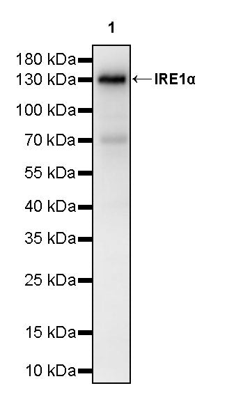
WB result of IRE1α Recombinant Rabbit mAb
Primary antibody: IRE1α Recombinant Rabbit mAb at 1/1000 dilution
Lane 1: COS-7 whole cell lysate 20 µg
Secondary antibody: Goat Anti- rabbit IgG, (H+L), HRP conjugated at 1/10000 dilution
Predicted MW: 110 kDa
Observed MW: 130 kDa