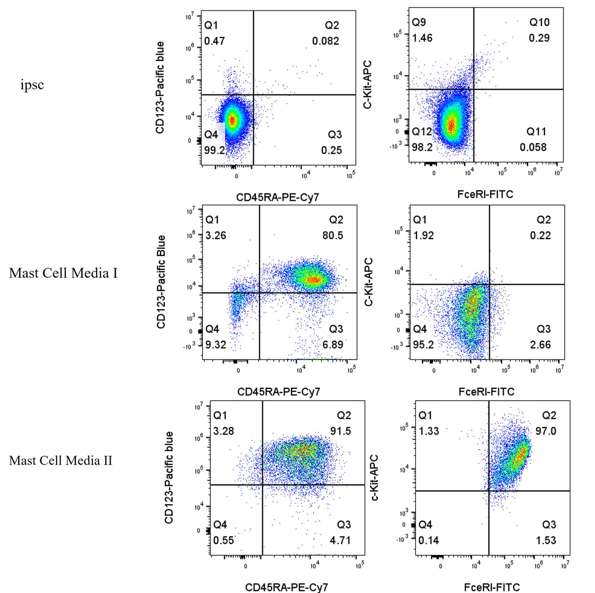
Flow cytometric phenotypic analysis of the iPSC-to-mast cell differentiation process. Undifferentiated iPSCs displayed a c‑Kit⁻/FcεRI⁻ phenotype. Following culture in Mast Cell Media I, the cells maintained negative expression for both c‑Kit and FcεRI. Notably, after 3 weeks of culture in Mast Cell Media II, the c‑Kit⁺/FcεRI⁺ double-positive population reached 97.0%, indicating the successful generation of mature mast cells (MCs).
hiPSC-to-Mast Cell Differentiation Cytokine Kit
hiPSC-to-Mast Cell Differentiation Cytokine Kit
Price:
Regular price
$435 USD
Regular price
Sale price
$435 USD
Unit price
per
For shipping services or bulk orders, you may request a quotation.
Secure checkout with

View full details
Product Details
Product Details
Product Specification
| Species | Human |
| Physical Appearance | Lyophilized powder |
| Reconstitution | Reconstitute at 0.1-1 mg/ml according to the size in ultrapure water after rapid centrifugation. |
| Stability & Storage |
·12 months from date of receipt, lyophilized powder stored at -20 to -80℃. · 3 months, -20 to -80℃ under sterile conditions after reconstitution. · 1 week, 2 to 8℃ under sterile conditions after reconstitution. · Please avoid repeated freeze-thaw cycles. |
Components
Component |
Reference dosage |
S Size(100ml system) |
M Size(1L system) |
FGF-basic(154aa) Protein, Human |
1-2 ng/mL |
5μg |
5μg |
IL-6 Protein, Human |
100 ng/mL |
10μg |
100μg |
SCF Protein,Human |
100 ng/mL |
10μg |
100μg |
FLT-3L Protein,Human |
25 ng/mL |
5μg |
5μg |
IL-3 Protein,Human |
5 ng/mL |
5μg |
5μg |
Protocol
Protocol for Differentiation of Human Induced Pluripotent Stem Cells (hiPSCs) into Mast Cells
1. Routine Culture of Human Induced Pluripotent Stem Cells (hiPSCs)
1.1 Seeding and Coating: Human induced pluripotent stem cells (hiPSCs) were seeded onto six-well plates pre-coated with Matrigel .
1.2 Medium and Culture Conditions: Cells were routinely cultured in mTeSR™ Plus complete medium at 37 °C in a 5% CO₂ humidified incubator. Medium changes and passaging were performed according to the growth status of the cells.
2. Induction of Embryoid Body (EB) Formation
2.1 Dissociation and Collection: When the iPSCs reached 70–90% confluence, the culture medium was aspirated, and the cells were gently rinsed once with 1 mL of PBS. After removing the PBS, 1 mL of mTeSR™ Plus dissociation reagent was added and incubated at room temperature for 30 seconds, The ReLeSR™ reagent was then aspirated.
2.2 Preparation of Cell Clumps: The plates were incubated at 37 °C in a 5% CO₂ incubator for 5 minutes. Then, 1 mL of mTeSR1 medium was added , and the culture surface was gently pipetted to disperse the cells into appropriately sized sheet-like or clump aggregates. Formation of a single-cell suspension should be avoided.
2.3 Suspension Culture and EB Induction: For cells from one T25 flask, the cell clumps were transferred to ultra-low attachment six-well plates at a 1:8 split ratio and cultured in EB induction medium (formula detailed below). The cells were cultured in suspension for 5 days, with medium changes performed every 2–3 days.
2.4 EB Induction Medium Composition: The EB induction medium consisted of DMEM/F12 basal medium, 20% KnockOut™ Serum Replacement (KSR), 1% Non-Essential Amino Acids (NEAA), 1% GlutaMax™, 0.1 mM 2-mercaptoethanol, and 1–2 ng/mL bFGF. (Note: 10 μM Y-27632 was supplemented during the first 24–48 hours of induction to enhance cell survival.)
3. Establishment and Culture of Myeloid Cell-Forming Complexes (MCFCs)
3.1 Transfer and Seeding: On day 6 of EB culture, EBs were collected using a cut pipette tip and seeded onto 6-well plates pre-coated with 0.1% gelatin at a density of 25–30 EBs per well.
3.2 MCFC Induction Culture: Mast Cell Medium I, as described below, was added to the cultures. The cells were then maintained for an additional 6–7 days to promote the formation of myeloid cell-forming complexes (MCFCs) and the generation of hematopoietic progenitor cells..
3.3 Mast Cell Media I Composition: Mast Cell Medium I consisted of APEL medium (STEMCELL, Cat. No. 05270), 100 ng/mL IL-6, 100 ng/mL SCF, 25 ng/mL Flt-3L, and 5 ng/mL IL-3.
4. Harvesting of Suspended Cells and Phenotypic Analysis
4.1 Observation of Release Phase: One week after EB seeding, the MCFCs began to gradually release suspended cells into the medium. The number of released suspended cells progressively increased during weeks 2–4 of culture.
4.2 Collection and Detection: To harvest the suspended cells, the side of the culture plate was gently tapped to facilitate the release of cells. The supernatant containing the suspended cells was collected. After centrifugation, the harvested cells were subjected to flow cytometry for the detection of surface markers (e.g., CD34, CD45, CD123, c-Kit, and FcεRI) to evaluate the induction of hematopoietic progenitors.
5. Maturation Induction of Mast Cells
5.1 Medium Change and Induction: The suspension cells released from MCFC into the culture medium were collected. After centrifugation and resuspension, the cells were transferred to Mast Cell Media II (MC Medium II; formula provided below) for maturation induction. The MCFC cultures were replenished with fresh Mast Cell Media I and maintained, and the suspension cells were collected once per week.
5.2 Maturation Culture: The cells were cultured in Mast Cell Media II for 2–3 weeks to promote terminal differentiation into mature mast cells. During the final week of culture, the cytokine concentration could be doubled to further facilitate mast cell maturation.
5.3 Quality Assessment: Following the maturation induction, cells were harvested and analyzed by flow cytometry to confirm mast cell identity by evaluating the expression of maturation markers, including c-Kit, FcεRI, CD45RA, and CD123.
5.4 Mast Cell Media II Composition: Mast Cell Medium II consisted of RPMI 1640 medium (containing GlutaMax™), 10% fetal bovine serum (FBS), 1% penicillin-streptomycin, 1 mM sodium pyruvate, 1% NEAA, 100 ng/mL SCF, and 100 ng/mL IL-6.
Guidelines
store in separate containers to reduce the number of freeze-thaw cycles.
Picture
Picture
Bioactivity