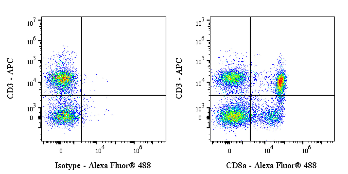
Flow cytometric analysis of SD rat splenocytes labelled Rat CD8a antibody at 1/500 dilution (1 μg) / (right panel) compared with a Mouse IgG1, κ Isotype Control / (left panel). Goat Anti-Mouse IgG Alexa Fluor® 488 was used as the secondary antibody. Then cells were stained with CD3 - APC antibody separately. Flow cytometry and data analysis were performed using Agilent NovoCyte Quanteon and FlowJo™ software.
Invivo anti-Rat CD8a Recombinant mAb
Invivo anti-Rat CD8a Recombinant mAb
Price:
Regular price
$175 USD
Regular price
Sale price
$175 USD
Unit price
per
For shipping services or bulk orders, you may request a quotation.
Secure checkout with

View full details
Product Details
Product Details
Product Specification
| Host | Mouse |
| Antigen | CD8a |
| Synonyms | T-cell surface glycoprotein CD8 alpha chain; CD8 antigen 32 kDa chain; OX-8 membrane antigen; Cd8a |
| Location | Cell membrane |
| Accession | P07725 |
| Clone Number | OX-8 |
| Antibody Type | Mouse mAb |
| Isotype | Mouse IgG1, κ |
| Isotype Control | Invivo mouse IgG1 isotype control |
| Application | FCM, in vivo CD8+ T cell depletion |
| Reactivity | Rt |
| Positive Sample | SD rat splenocytes |
| Purification | Protein G |
| Concentration | 5 mg/ml |
| Purity | >95%(Determined by SDS-PAGE) |
| Endotoxin | <1EU/mg |
| Conjugation | Unconjugated |
| Physical Appearance | Liquid |
| Storage Buffer | PBS pH7.4, containing no preservative |
| Stability & Storage | 2 to 8 °C for 2 weeks under sterile conditions; |
Dilution
| application | dilution | species |
| FCM | 1:500 | Rt |
Background
CD8A, also known as T-cell surface glycoprotein CD8 alpha chain, is a protein encoded by the CD8A gene. It is a cell surface glycoprotein found on most cytotoxic T lymphocytes, mediating efficient cell-cell interactions within the immune system. The CD8 protein acts as a coreceptor with the T-cell receptor on T lymphocytes to recognize antigens displayed by antigen-presenting cells in the context of class I MHC molecules. The functional coreceptor can be either a homodimer composed of two alpha chains or a heterodimer composed of one alpha and one beta chain. CD8A has significant homology to the variable domain of immunoglobulin light chains. It plays an essential role in the immune response, enabling cytotoxic T lymphocytes to recognize and eliminate infected cells and tumor cells.
Picture
Picture
FC