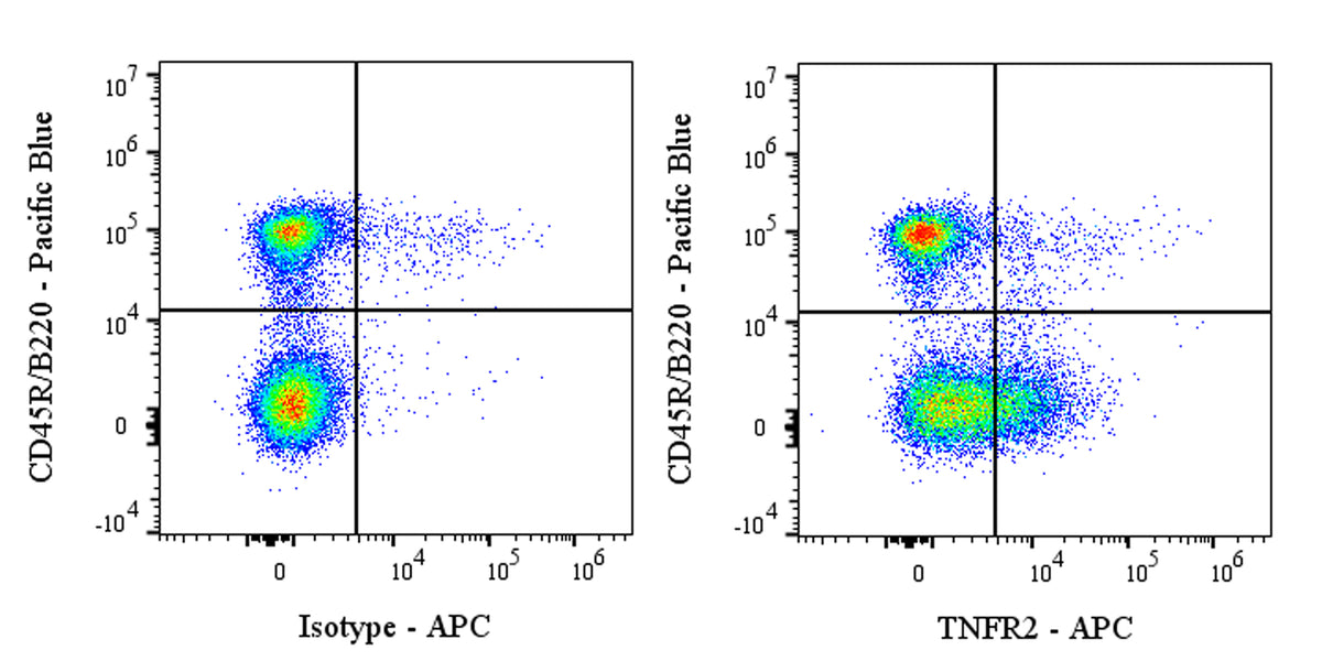
Flow cytometric analysis of C57BL/6 mouse splenocytes labeled with Anti-Mouse TNFR2 (CD120b) antibody at 1/500 dilution (1 μg) / (right panel) compared with an Armenian hamster IgG Isotype Control / (left panel). Biotin‑conjugated Anti‑Armenian hamster IgG was used as the secondary antibody, followed by Sav-APC. Then cells were stained with SDT CD45R/B220 - Pacific Blue antibody (S0B5627) separately. Flow cytometry and data analysis were performed using Agilent NovoCyte Quanteon and FlowJo™ software.
Invivo Anti-Mouse TNFR2 (CD120b) Recombinant mAb
Invivo Anti-Mouse TNFR2 (CD120b) Recombinant mAb
Price:
Regular price
$175 USD
Regular price
Sale price
$175 USD
Unit price
per
For shipping services or bulk orders, you may request a quotation.
Secure checkout with

View full details
Product Details
Product Details
Product Specification
| Host | Armenian hamster |
| Antigen | TNFR2 (CD120b) |
| Location | Membrane |
| Accession | P25119 |
| Clone Number | S-5573 |
| Antibody Type | Recombinant mAb |
| Isotype | Armenian hamster IgG |
| Isotype Control | S0B7062 |
| Application | FCM, in vivo blocking, in vitro blocking |
| Reactivity | Ms |
| Positive Sample | C57BL/6 mouse splenocytes |
| Purification | Protein G |
| Concentration | 5 mg/ml |
| Purity | >95% (Determined by SDS-PAGE) |
| Endotoxin | <1EU/mg |
| Conjugation | Unconjugated |
| Physical Appearance | Liquid |
| Storage Buffer | PBS pH7.4, containing no preservative |
| Stability & Storage | 2 to 8 °C for 2 weeks under sterile conditions; |
Dilution
| application | dilution | species |
| FCM | 1:500 | Ms |
Background
TNFR2, or tumor necrosis factor receptor 2 (encoded by the TNFRSF1B gene), is a type I transmembrane glycoprotein of approximately 75 kDa belonging to the TNF receptor superfamily. It is primarily expressed on the surface of immune cells (such as Treg cells, myeloid-derived suppressor cells or MDSCs, endothelial cells, and certain tumor cells). Unlike the ubiquitously expressed TNFR1, TNFR2 exhibits higher affinity for its ligand TNF-α but is only efficiently activated when high concentrations of TNF-α are present in specific microenvironments (such as inflammatory or tumor microenvironments). Functionally, TNFR2 not only participates in classical pro-inflammatory signaling (by recruiting adaptor proteins such as TRAF2 to activate NF-κB and PI3K/AKT pathways, promoting cell survival, proliferation, and angiogenesis) but also plays a "double-edged sword" role in immune regulation: on one hand, TNFR2 is a critical receptor for maintaining Treg cell stability and suppressive function, as its signaling promotes Treg expansion and enhances their immunosuppressive capacity; on the other hand, within the tumor microenvironment, TNFR2 highly expressed on tumor cells—upon binding TNF-α—promotes tumor invasion and metastasis through the NF-κB pathway, while TNFR2 on tumor-infiltrating Tregs exacerbates local immunosuppression, and TNFR2 on MDSCs promotes their accumulation and suppressive activity. Furthermore, TNFR2 has been recognized as a "survival switch" for tumor cells to evade T-cell killing—by inducing anti-apoptotic protein expression to resist TNF-mediated cytotoxicity. Given its dual pivotal roles in immunosuppression and tumor survival, TNFR2 has emerged as a prominent novel immunotherapy target following PD-1/PD-L1, with various anti-TNFR2 agonistic antibodies (for activating Tregs to treat autoimmune diseases) and antagonistic antibodies (for blocking tumor immunosuppression) currently in preclinical and early-stage clinical development, aiming to achieve precise regulation of immune responses for therapeutic purposes.
Picture
Picture
FC