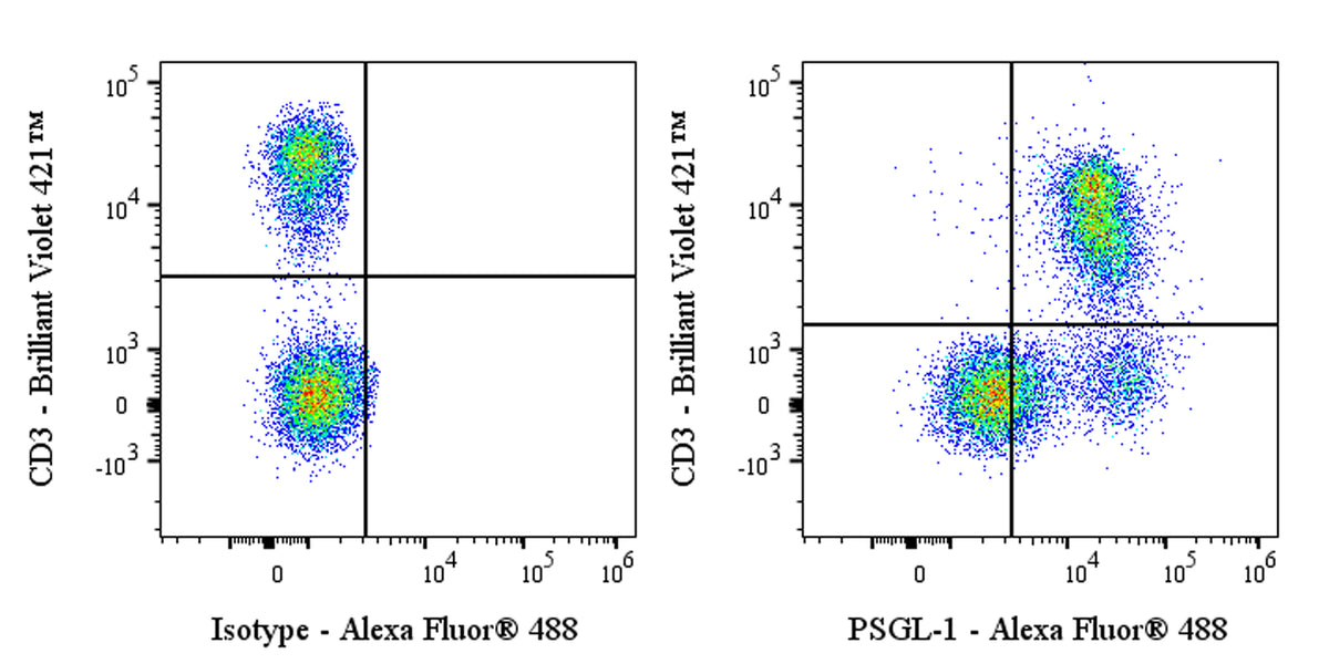
Flow cytometric analysis of C57BL/6 mouse splenocytes labeled with Anti-Mouse PSGL-1 (CD162) antibody at 1/500 dilution (1 μg) / (right panel) compared with a Rat IgG1, κ Isotype Control / (left panel). Goat Anti-Rat IgG Alexa Fluor® 488 was used as the secondary antibody. Then cells were stained with CD3 - Brilliant Violet 421™ antibody separately. Flow cytometry and data analysis were performed using Agilent NovoCyte Quanteon and FlowJo™ software.
Invivo Anti-Mouse PSGL-1 (CD162) Recombinant mAb
Invivo Anti-Mouse PSGL-1 (CD162) Recombinant mAb
Price:
Regular price
$175 USD
Regular price
Sale price
$175 USD
Unit price
per
For shipping services or bulk orders, you may request a quotation.
Secure checkout with

View full details
Product Details
Product Details
Product Specification
| Host | Rat |
| Antigen | PSGL-1 (CD162) |
| Location | Cell membrane |
| Accession | Q62170 |
| Clone Number | S-5511 |
| Antibody Type | Rat mAb |
| Isotype Control | S0B7122 |
| Application | FCM, in vivo blocking |
| Reactivity | Ms |
| Positive Sample | C57BL/6 mouse splenocytes |
| Purification | Protein G |
| Concentration | 5 mg/ml |
| Purity | >95% (Determined by SDS-PAGE) |
| Endotoxin | <1EU/mg |
| Conjugation | Unconjugated |
| Physical Appearance | Liquid |
| Storage Buffer | PBS pH7.4, containing no preservative |
| Stability & Storage | 2 to 8 °C for 2 weeks under sterile conditions; |
Dilution
| application | dilution | species |
| FCM | 1:500 | Ms |
Background
PSGL-1, or P-selectin glycoprotein ligand-1, is a type I transmembrane glycoprotein encoded by the SELPLG gene on human chromosome 12 and belongs to the mucin family. It is primarily expressed on the surface of leukocytes, including neutrophils, monocytes, T cells, and B cells. Its molecular structure features a prominent N-terminal extracellular region containing multiple mucin-like repeats rich in serine, threonine, and proline, which are heavily modified with O-glycans and sialyl Lewis X (sLeX) glycosylation—these glycosylations form the structural basis for PSGL-1 binding to its ligands, P-selectin, E-selectin, and L-selectin. The most classic physiological function of PSGL-1 is to serve as the cognate counter-receptor for P-selectin, mediating the initial capture, rolling, and slow adhesion of leukocytes on endothelial cell surfaces under flow shear stress at sites of inflammation—this represents the first step in leukocyte extravasation into inflamed tissues and is a critical process in immune cell homing and migration. However, the functions of PSGL-1 extend far beyond those of a mere adhesion molecule: accumulating evidence in recent years has established PSGL-1 as an inhibitory receptor in T-cell receptor (TCR) signaling. When cross-linked with specific endogenous ligands (such as certain chemokines or extracellular matrix proteins), PSGL-1 can downregulate signaling events like ERK phosphorylation, thereby suppressing T-cell proliferation, production of effector cytokines such as IFN-γ, and secretion of IL-2—thus negatively regulating T-cell activation. Furthermore, PSGL-1 plays complex and important roles in tumor immunity: on one hand, within the tumor microenvironment, P-selectin highly expressed by tumor cells or stromal cells can mediate platelet-leukocyte aggregation through PSGL-1, forming immune shielding barriers that promote tumor immune evasion. On the other hand, high expression of PSGL-1 on tumor-infiltrating lymphocytes (TILs) is closely associated with T-cell exhaustion, and blocking PSGL-1 has been shown to rejuvenate the anti-tumor function of CD8⁺ T cells, demonstrating synergistic potential with PD-1/PD-L1 blockade in preclinical models. Therefore, PSGL-1 has emerged as a novel and promising target in cancer immunotherapy, with anti-PSGL-1 monoclonal antibodies currently in early-stage clinical development, offering hope for new therapeutic avenues for cancer patients.
Picture
Picture
FC