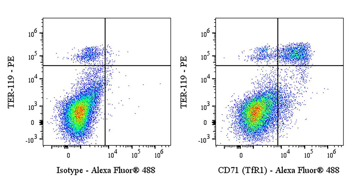
Flow cytometric analysis of BALB/c mouse bone marrow labeled with Anti-Mouse CD71 at 1/500 dilution (1 μg) / (right panel) compared with a Mouse IgG2a, κ Isotype Control / (left panel). Goat Anti- Mouse IgG Alexa Fluor® 488 was used as the secondary antibody. Then cells were stained with TER-119 – PE antibody separately. Flow cytometry and data analysis were performed using Agilent NovoCyte Quanteon and FlowJo™ software.
Invivo Anti-Mouse CD71 (TfR1) Recombinant mAb
Invivo Anti-Mouse CD71 (TfR1) Recombinant mAb
Price:
Regular price
$175 USD
Regular price
Sale price
$175 USD
Unit price
per
For shipping services or bulk orders, you may request a quotation.
Secure checkout with

View full details
Product Details
Product Details
Product Specification
| Host | Mouse |
| Antigen | CD71 (TfR1) |
| Synonyms | Transferrin receptor protein 1; TR; TfR; Trfr; Tfrc |
| Location | Cell membrane |
| Accession | Q62351 |
| Clone Number | S-5107 |
| Antibody Type | Mouse mAb |
| Isotype | Mouse IgG2a, κ |
| Isotype Control | S0B0787 |
| Application | FCM, in vivo cell specific depletion |
| Reactivity | Ms |
| Positive Sample | BALB/c mouse bone marrow |
| Purification | Protein A |
| Concentration | 5 mg/ml |
| Purity | >95% (Determined by SDS-PAGE) |
| Endotoxin | <1EU/mg |
| Conjugation | Unconjugated |
| Physical Appearance | Liquid |
| Storage Buffer | PBS pH7.4, containing no preservative |
| Stability & Storage | 2 to 8 °C for 2 weeks under sterile conditions; |
Dilution
| application | dilution | species |
| FCM | 1:500 | Ms |
Background
CD71, also known as the transferrin receptor 1 (TfR1), is a type II transmembrane glycoprotein that plays a crucial role in cellular iron uptake by binding to transferrin, which facilitates iron transport into the cell. This receptor is highly expressed on rapidly proliferating cells, including erythroid precursors, activated immune cells, and cancer cells, as these cells require significant iron for DNA synthesis and metabolic processes. In clinical contexts, CD71 is used as a marker for identifying immature red blood cells (such as proerythroblasts and reticulocytes) and is also associated with aggressive tumor growth in various cancers, including pancreatic and colon cancers. Additionally, studies have shown that CD71 expression on neutrophils can be linked to poor prognosis in pancreatic ductal adenocarcinoma (PDAC) patients, indicating its potential as a prognostic biomarker.
Picture
Picture
FC